
دسته: Gamacmera- Spect
فیلتر محصولات
نمایش 1–16 از 25 نتیجه
-

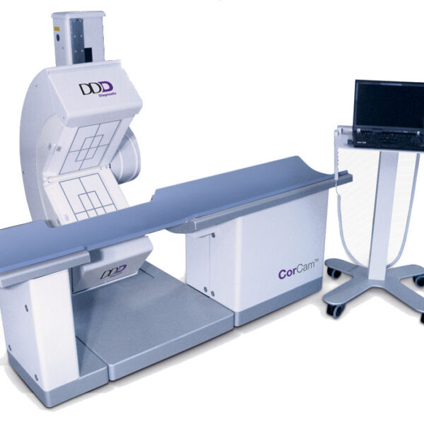
DDD CorCam- Dedicated Cardiac Gamma Camera
اطلاعات بیشتر -
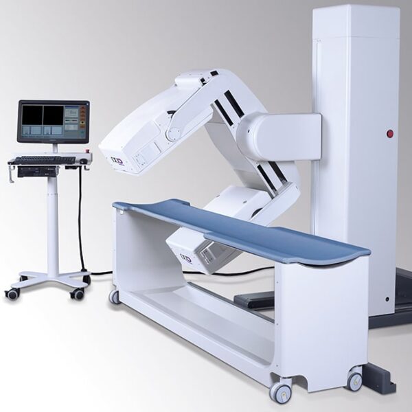
DDD- QuantumCam
اطلاعات بیشتر -

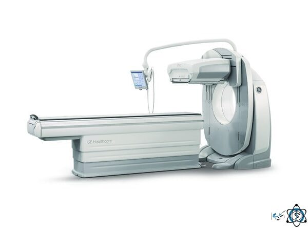
GE – BRIVO- NM615 – Single Head
اطلاعات بیشتر -

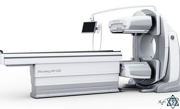
GE- Discovery NM 630- Dual Head Gamma Camera
اطلاعات بیشتر -
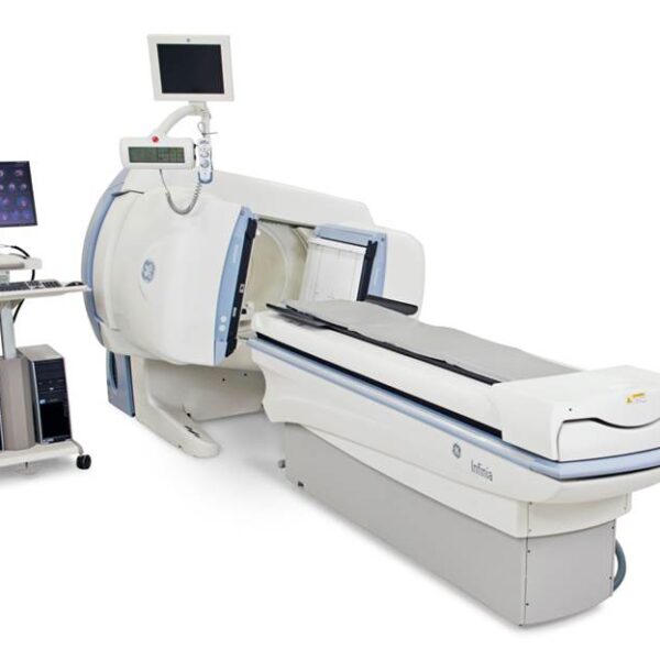
GE- Infinia
اطلاعات بیشتر -

MultiSPECT 2 Siemens
اطلاعات بیشتر -

Philips (ADAC) Vertex
اطلاعات بیشتر -

Philips (ADAC)- Forte
اطلاعات بیشتر -

Philips (ADAC)- Genesys- Dual Head
اطلاعات بیشتر -

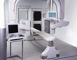
Philips (ADAC)- SKYLight
اطلاعات بیشتر -
Philips- BrightView X
اطلاعات بیشتر -

Picker- PRISM 2000
اطلاعات بیشتر -

Picker- PRISM 3000
اطلاعات بیشتر -
Siemens ( MiE )- DIACAM SCINTRON- Single Head
اطلاعات بیشتر -
Siemens ( MiE )- ECAM Scintron
اطلاعات بیشتر -

Siemens c cam
اطلاعات بیشتر

Gamacmera- Spect
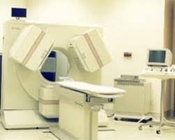
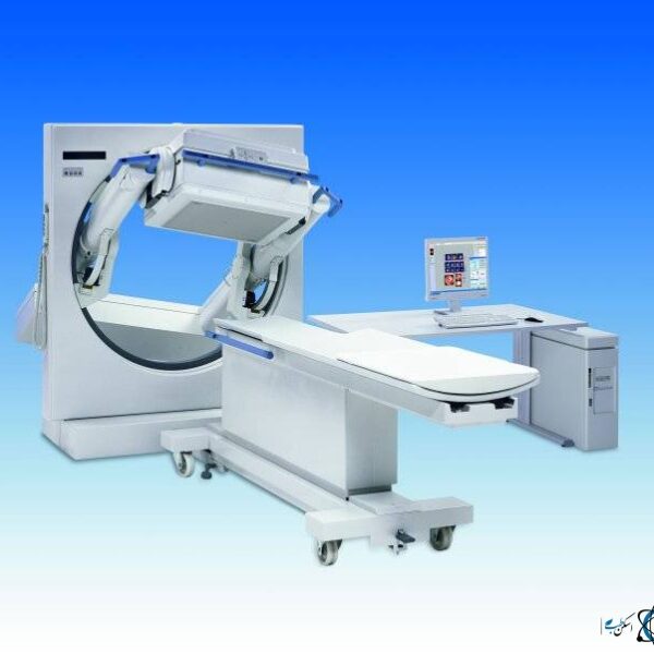
Gamacamara – Spect is one of the medical imaging devices that has come to the aid of doctors in diagnosing various diseases. Single Photon Emission Computed Tomography (SPECT) for decades in Nuclear Medicine It is used for cardiovascular scans, bone scans, kidney scans, etc. to provide 3D images of radiographic radiation distribution.
This SPECT camera detects radiation emitted by injectable radioisotopes and forms an image of their distribution in the body. Generally, in radioisotope imaging methods or nuclear medicine, because different tissues of the body absorb different amounts of radioactive matter depending on the type of scan, the size, shape and position of a limb can be investigated relative to the surrounding environment of that organ.
The difference between nuclear medicine imaging and other medical imaging methods is in providing pathological images compared to anatomical images in other medical imaging methods.
Although SPECT data in general are superior to planar data, sometimes the use of SPECT data has some weaknesses due to its inability to accurate anatomical localization of an anomaly. Many of these limitations can be compensated by combining spect imaging with an anatomical imaging method such as computed tomography (CT). In the Spect/CT topic, we will also familiarize you with the SPECT-CT hybrid imaging method.
What is Radiodaro and what is its application in nuclear medicine:
Radiopharmaceuticals or radioactive substances are substances that are often used in liquid form and with specific activity in various topics, especially medicine. In most cases, radioactive material enters the body by intravenous injection and after accumulation in the tissue, the resulting radiation is imaging.
Two issues should be considered in the use of radiopharmaceves in the nuclear medicine sector. These two issues are the half-life of the radioactive material and its energy. The half-life of the radioactive substance used should not be long enough for the patient and those around him to be exposed to radiation for a long time, and on the other hand, it should not be short enough to significantly reduce its activity before or during imaging. Radioactive material energy is also important for its detection in the system.
The sources used in gamma cameras are gamma-ray diffuse isotopes, the most common of which is technium. Technium has a half-life of 6 hours and is removed from the patient’s body within one day (its physiological excretion is appropriate). This radioisotope has good absorption and has KeV140 energy. A radioactive drug contains certain chemicals that are marked with a radionuclide.
Overall structure and performance of Gamma Camra:
The basis of the gamma-gamma device, or gamma and spect camera (SPECT), is the ignition count that is caused by the collision of gamma rays to a plate called a crystal. Photons emitted from the body are absorbed or dispersed due to the presence of different tissues and barriers in their way. Any of this can change its energy or divert photons from its path.
However, due to these phenomena, we have some unhelpful and scattered radiations that reduce the accuracy of imaging. To remove these beams, the climator is used (which varies depending on the isotope energy used by the type of collimators used), to prevent such photons from entering the ashkasaz and prevent a significant number of diagonal and scattered beams from hitting the crystal.
The use of collimators causes parallelization of the beams and photons hit the crystal, causing interactions and the burning action or ignition occurs. Photo-Multiplier Tube-PMT is located behind the crystal collecting optical pulses and according to the electronic circuit connected to PMTs and pulses reached to each PMT, the location is performed and the original pulse value with location (x,y) is provided to the reconstruction algorithm as input.
After the reconstruction process, an image is obtained that can be seen on a display in a color with different intensities, which is actually the image of the member or organ in question.
Components of gamma-merma machine:
Gamma camera or more comprehensive SPECT device can be divided into four main parts. This device includes gantry, detector or detector, console and bed.
Gantry Gamacomera Device:
Gantry is the trunk of the device, which is the mobile part of the device in most of the devices in the medical equipment market, and the location of the detector is gamma-amra device.
Gamacama device detector and its components:
The gamma-amara device detector, as its name suggests, plays the role of detecting radiation output from the patient’s body or received. The gamma-amara detector is composed of components such as climators, detectors or crystals, photomalated play tubes (PMT) and electrical circuits. Gamacma device detector is the most important and at the same time the most expensive part of the device.
The gamma ray detector absorbs itself and produces itself to the electrical signal in accordance with the same adsorption locations and sends these signals to the console of the gamma-gamma device.
In the following, we will briefly explain the different components of the gamma-gamma device detector.
1. Climator:
Climators are usually composed of very large pieces of lead that have apertures that are parallel to each other. Collimators are made to pass only beams that move parallel to the apertures.
The purpose of using collimator in gamma-gamma device is to remove radiation other than the main beams in the tissue site. For example, diagonal and scattered beams that come from collisions with the patient’s body tissue or the device bed to the detector.
2. Crystal:
The role of crystals in the detector of gamma system is to convert gamma rays into optical photons and transfer to PMTs of gamma-gamma machine. The crystals used in the gamma-mera machine have different types, usually made of sodium iodide (NaI). That brings a small amount of tullym impurities. The object is sensitive to light and radiates luminous photons by absorbing gamma rays. These photons have a wavelength of about 410 nm.
3. Photomolethylier Tube (PMT):
PMT or so-called PMT Tube includes: Photocatad is a high voltage power supply (electron amplifier) and finally the output part. When photons are absorbed by crystals and a luminous spark is created, each tube produces a flow output pulse. Therefore, PPM Tube acts like a visible light converter to electrical current.
The pulse amplitude of each tube is directly proportional to the amount of light that the photocatalyst has received, the resulting light in the photomaholtic photocatalytic layer is converted into a number of low-energy electrons. Then, the electrons emitted from photocatade accelerate from one dynaud to the next with an overall potential difference of about 2000 volts during photomoletti pleir.
By hitting each electron to the dynaudal surface, two or three electrons are irradiated from it, thereby increasing the amplified gain. Finally, the photomolethy-pleir output current can be applied to the amplifier circuit, to be used in power measurement devices, scales or screens.
Those light bulbs near the point of light producing the largest pulses and those that are far from it produce small signs, so each tube produces an electrical pulse proportional to the amount of proximity to the spark. In some cases, the output pulses are so small that they are lost in conventional electrical jamming of the tube, and therefore have no visual applications.