Preparations before pet scan
Pet Scan and its introduction:
Pet Scan
is a diagnostic imaging tool. PET imaging is a process by which biological functions of the body are able to be imaging, and this is very attractive for doctors. Using a small amount of a biological marker that is the same as radiopharmaceno, Pen scans can evaluate areas of abnormal glucose metabolism in the body.

The high use of glucose or sugar in cancer is based on an important discovery made in 1921 by Otto Warburg, when he was studying the metabolism of cancer cells. He found that breathing oxygen in normal body cells in cancer cells was replaced by sugar fermentation. The body’s natural cells meet their energy needs through oxygen respiration, while cancer cells supply their energy needs largely through fermentation.
From a biophysical and biochemical perspective, this difference between normal and cancerous cells is remarkable. Oxygen gas is an energy source in flora and fauna. In cancer cells, this process is replaced by an energy-generating reaction from the lowest living forms, namely glucose fermentation. In the 1970s the Brookhaven Group produced 18F-FDG isotopic radio that can carry sugar. In 1975 the first pet scan device was built by Michael E. Phelps, Phelps, PhD, chief of the Department of Molecular Pharmacology and Medicine at the David Geffen School of Medicine at UCLA.
The pen scan device allowed us to conduct the first clinical studies in the field of neurology, cardiology and oncology by tracking the absorption of radioactive isotope used in PET imaging to determine how body organs use sugar. Research continued to progress, pet scan devices got better and eventually imaging studies were carried out. Pet scans are incredibly useful because cancer cells, naturally, are rapidly dividing.
After 90 hours of cell division, a cancer cell can be converted into 32,768 thousand cells. A 1cm tumor contains 10 billion cancer cells. This rapid cell division requires a lot of fuel and it is the sugar fuel in our bodies. However, suddenly don’t throw away all the foods containing sugar. Keep reading because we will address this specifically and your concerns below!
During the 1980s and 1990s the quality of pet scans improved and the scans became more useful and specific. FDG, a sugar-based isotope, decomposes quickly, so linear accelerators are needed in place to produce FDG. Since the 1980s, the number of centers using pet-scanning devices has increased.
One of the major advances in imaging technology was the ability to integrate CT and PET devices into one device and put images one over the other. CT scan shows anatomical structures but is not effective enough in determining the characteristics of structures. Pet scans show whether there is a strong absorption of sugar and therefore it is useful in detecting the presence of most cancers. Pet/CT produces an image of anatomy combined with functional images of the use of sugar by cells.
Pet scan imaging helps determine where the primary tumor is located. For example, it can help determine whether cancer can be surgically picked up or whether lesions are outside the scope of surgical intervention. It is also useful in determining the rate of cancer progression. Today, pet scan has become more important as a tool in assessing whether a treatment protocol works well.
As a result, patients can stop ineffective treatments more quickly and move towards more effective treatment for their cancer. This is a vital decision for patients and doctors as it helps prevent unnecessary exposure to the side effects of ineffective treatments. By preventing unnecessary, and often very costly, treatments, it saves money, and most importantly, it allows the cancer specialist to push the patient into more effective treatment.
Pet Scan is useful for helping to diagnose:
- Oncology – to determine the location of tumors and the rate of tumor growth
- Neurology – to help diagnose Alzheimer’s, dementia, stroke, brain tumors and seizures
- Cardiology – to determine the viability of the heart muscle
How to do pet scan
At the beginning of the pet scan procedure, you sit in a chair and a small amount of radioactive substance called FDG is injected into the vein of your arm. This injection doesn’t make you feel any different.
You will be asked to sit quietly for a while, usually about 30 to 60 minutes. During this time you have to sit and relax and you can’t get up and walk, talk to friends or study. This expectation will allow the injected radioactive substance to move throughout your body.
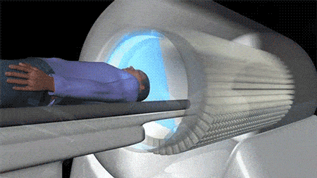
You will then be asked to lie on the bed of the pet scan device. The pet scan machine has a larger opening than an MRI and does not produce any sound. A pet scan imaging usually takes about an hour to complete.
Preparations before pet scan
In the following article, from the scan of medicine and articles about pet scans, pre-pet scan preparations can be divided into the previous day’s diet until the scan.
Diet Before Pet Scan

When planning to do a pet scan, it is important to keep in mind that your doctor will ask you to observe a pre-pet scan diet up to 12 hours before your pet scan turn. A typical pet scan diet is as follows:
12 hours before pet scan:
You will be asked to have a low-carb diet. Foods that can be consumed include: Meat, stiff cheese, eggs, butter and vegetables without starch.
Some foods that are not allowed include: Cereals, pastas, milk, bread and other sugars.
If your pet scan turn is booked in the late afternoon scheduled, you can have breakfast before scanning. You should also have a low-carb diet.
6 hours before pet scan:
For all pet scan regimens, you’ll be asked not to eat 6 hours before pet scans. However, you may be allowed to continue drinking water.
You will also be asked to bring copies of recent CT scans or previous MRI. It should be noted that if you have already taken a CT scan or an MRI turn from a medical scan, it is likely that their images will be available in your profile and you will no longer need to take these photos.
After waiting time and injecting the radioactive substance, you will be taken to the scan room for pet scan and you can go home after the scan is finished and some rest.
What to eat the day before pet scan?
Stay on a diet with very low carbs and no sugar the day before pet scan. Avoid eating or drinking caffeinated products (chocolate, soft drinks, tea, coffee) 24 hours before pet scan.
Why do you need a special diet before pet scan?
The radiodrug used in pet imaging is fluorine 18 fluorodoxic glucose, or 18F-FDG, a glucose transporter. This radioisotope goes to any metabolically active area in the body. If glucose levels rise from the food or drink that the patient has consumed before pet scans, insulin levels increase. As a result, this radiodrug, which is accompanied by a lot of light, does not accumulate in the tissues of cancer cells and pet scan image does not have enough readability for the doctor.
Final Word
In this article, we will use the scan of medicine to the preparations before pet scan, which we hope will help you to do pet scans.
Nowadays, the use of pet scan despite the increase in cancer cases, especially in Iran, has increased, and on the other hand, taking the turn of pet scan is equally challenging. We have created a solution in the scan of medicine to solve this problem. You can get a free pet scan turn in the medical scan. You can also take turns from your nearby pet scan. To do this, you need to take a free pet scan from the medical scan page.


