Vaginal ultrasound
Vaginal ultrasound
What is vaginal ultrasound or uterine ultrasound?
Ultrasound or ultrasound is a term used for high-frequency sound waves. Ultrasound imaging uses these sound waves to produce an image from inside the body that is visible on the screen. Ultrasound is performed by a trained health professional (radiologist or sonologist). Vaginal ultrasound or sonovaginal is the process of pelvic examination in women. This type of imaging helps your doctor carefully examine any abnormalities inside the uterus, cervix (cervix), endometrium (inner lining of the uterus), fallopian tubes, ovaries, bladder or pelvic cavity.
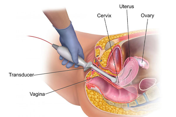
Vaginal ultrasound helps us see pelvic organs from inside the vagina using a smooth, thin, handheld probe called transducer, ultrasound probe or transducer. Vaginal ultrasound is different from abdominal ultrasound. In abdominal ultrasound, it uses a hot water-based transparent gel applied to the skin of the abdomen and the transducer moves slowly in the pelvic area.
All transducers or ultrasound probes transmit high-frequency sound waves and these waves are reflected from soft tissue, structures or different parts of the body in different ways. These sound waves are transformed into electrical impulses that display a motion picture on the screen. This can also be provided for vaginal ultrasound.
Ultrasound has many advantages. First and foremost, it is painless and risky, which does not include ionized radiation. There is no injection in the ultrasound unless your doctor has specifically requested it. High-frequency sound waves allow images to be produced in very high detail. These images can also look at the tiniest parts of the body. At the time of vaginal ultrasound imaging, a specialist doctor will be by your side, and you have the opportunity to share any concerns you have.
When is vaginal ultrasound performed?
The appropriate time for a vaginal ultrasound is requested if you have symptoms of pelvic pain or abnormal bleeding, or to check for fibroids (muscle tumors of the uterus), polyps (thickened areas of the inner lining of the uterus), cysts or ovarian tumors, infertility, this scan is requested by your doctor. Or your doctor may prescribe an abdominal ultrasound scan using a smooth handheld transducer on the outer part of the body, in the lower abdomen or sono vaginal area, to assess the early pregnancy periods.
Vaginal ultrasound provides much more detailed images of pelvic organs, especially the uterus (including fallopian tubes and ovaries) compared to pelvic ultrasound scans of the lower abdomen.
How to prepare for uterine or vaginal ultrasound
There is no pre-preparation for uterine ultrasound
. Before performing the scan, you will be asked to go to the toilet and empty your bladder.
If you are on a monthly period, scans can still be performed and are often an advantage in assessing some of the problems women have. If you are using sanitary pads, it should be removed.
Vaginal ultrasound is an invasive personal examination, so you may be asked to sign a consent letter before the test. In any case, the patient’s dignity and privacy are protected during the examination.
The examination is carried out by sonographers who may be male or female. Upon arrival at the radiology center, if you are not comfortable with an ultrasound by the man, you should notify the reception staff and ask a female sonographer to perform the examination. If your female doctor is unavailable, your scan turn may change. If you have an abdominal ultrasound, you should drink two to three glasses of water 30 minutes before the test to fill the bladder. It’s best to wear a comfortable dress that has easy access to the lower part of your body.
How to do a vaginal sono?
How to perform a vaginal ultrasound after discharge of the bladder is so that you are asked to remove your clothes from the waist down. You will then be asked to lie on the scan bed. In general, a veneer is thrown on the scanned limb to cover you. You will be asked to bend your legs and a special transducer will enter the vagina. The transducer does not have a large diameter and is not annoying, and is specifically a form that is easily placed in the vagina. A protective coating is placed on the transducer and the hot lubricant gel is rubbed on it for ease of placement. It moves slowly inside the pelvis and images are taken. Your doctor may press your underbelly by hand to bring some of the pelvic organs closer to the vaginal probe to get better images.

The examination is done in “real time” meaning that the images you see on the screen show the inside of your pelvic area (lower abdomen). Still photos are also taken during the examination. At the end of the scan, the probe is completely sterile and cleaned.
Get a scan turn
The Specialized Website of Scan Teb has set up a panel for the convenience of patients to obtain medical scans and medical imaging so that patients can easily choose the date of the scan and their medical imaging center. Through the link below, you can choose the scan turn, the type of scan, the city you want to scan and the date and time of your scan. You can use the Scan Teb website to get an
ultrasound appointment
.


