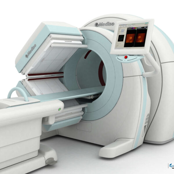
SPECT – CT
History of SPECT CT:
Although the SPECT-CT imaging method was first introduced in 1990, its commercial use has expanded since the production of PET-CT. That is, the advantages of PET CT were first identified for physicians and as a result, many doctors turned to SPECT-CT.
Many of the properties of PET-CT and SPECT-CT are similar. For example, while anatomical imaging techniques allow for accurate diagnosis and location of morphological abnormalities, nuclear medicine studies reflect the pathophysiological status of the disease.
To put it simply, the CT scan shows the physical shape of the body tissue in detail but does not provide information about how that particular tissue works. While the SPECT image can show how that organ works. To illustrate، we provide an example. Suppose a doctor wants to have an image of the patient’s kidney tissue. With a CT scan, the doctor can see the exact location of the kidney in detail.
This kidney tissue may appear perfectly healthy on the surface, but there may be severe kidney failure due to some problems. No doctor can tell how kidney tissue works from a CT scan. That’s where Spect’s leg opens up to the field.
A kidney SPECT image can tell the doctor which parts of the kidney are active and which parts are failing. As a result, these two imaging techniques together can cover each other’s weaknesses well, and doctors can use their strengths.
Another important and similar property of SPECT CT and PET CT is attenuation correction. Correction of attenuation in medical images and especially nuclear medicine is very important and improves the accuracy of images.
The first attempts to build Spect CT were probably made by Hoggawa and his colleagues. They tried to develop a system capable of simultaneously carrying out CT and SPECT studies, which paved the way for the development of hybrid SPECT-CT systems for clinical use.
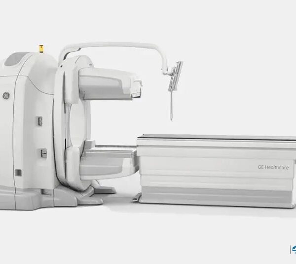
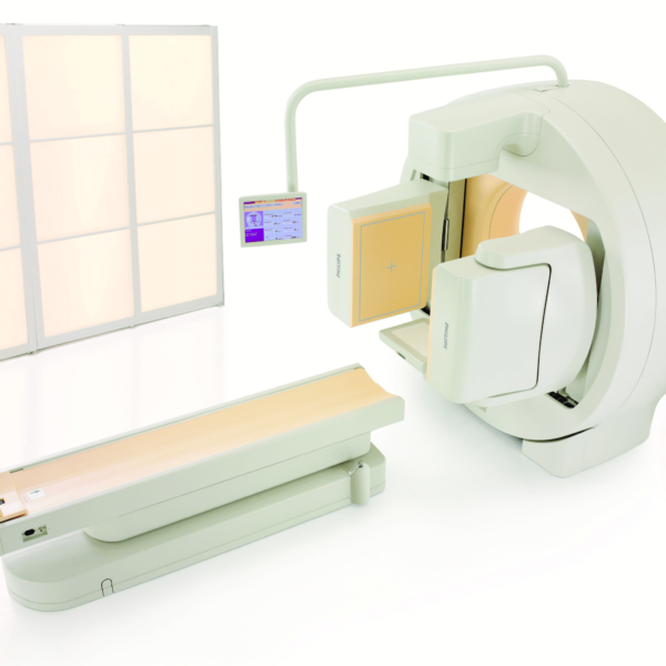
The first commercial system called Hawkeye was developed by General Electric in 1999. After that, other manufacturers decided to bring commercial SPECT-CT devices to market. For example, Philips’ Precedent device and Siemens Symbia are examples of successful models.
It’s time to talk a little bit about Spectac-CT images. To correct attenuation, it is necessary to map attenuation from the spatial distribution of attenuation coefficients for each patient. The attenuation map is then used by a repetitive reconstruction algorithm to perform attenuation correction for the given release data.
In the past, this attenuation correction was done with radionuclide-based transport images, but this method was rarely used clinically in practice. Now, CT-based attenuation correction has become the standard for PET and is emerging rapidly as the SPECT standard.