دسته: MRI
Magnetic resonance imaging or MRI
If you haven’t stepped into diagnostic treatment centers to shoot your body, you’ve still heard of an MRI. MRI is no longer a technique, but a term and concept of medical diagnosis in the public mind. So if you’ve only ever had a name in your mind from this imaging technique and you’re going to improve your information about physics and how the MRI machine works in simple terms, we suggest you don’t miss this article.
If we simply intend to define mri generalities, we can say that magnetic resonance imaging, or MRI, allows us to view the human body in amazing detail using magnets and radio waves. The first MRI scanner used to imaging the human body was developed in New York in 1977. Since then, this technology has been greatly enhanced and MRI is now frequently used by all physicians in the world as a standard and reliable imaging. Perhaps one of the key advantages of MRI imaging is that MRI does not involve high-risk radiation (such as X-rays or CT scans) and has no invasive effects on the body.
The MRI scanner is essentially a giant magnet. Magnet strength is measured in a standard unit called Tesla (T). Most MRI scanners used in medical research hospitals and clinics are 1.5 or 3 Teslas. To clarify Tesla’s large scale, it’s not bad to know that Earth’s magnetic field is about 0.00006 Tesla!!!
The real question is, what is the use of this ultra-strong magnetic field? To answer this question, you should know that all this magnetic force is spent absorbing water molecules into the body’s tissues.
So we have to see what these hydrogen atoms are capable of that are so vital in imaging. If we examine the hydrogen atom more carefully, we see that it contains a central nucleus containing a single positive charge called proton. These protons orbit around their axis with the magnetic poles of north and south, and each hydrogen proton is spinning like a tiny magnet that rotates on its axis. This rotational motion is known as Precession. At any moment in time, all the billions of hydrogen protons in our bodies are in random positions and revolving around their axes.
However, these random situations change altogether when placed in a very strong magnetic field. It’s just like when a compass needle aligns with the Earth’s magnetic field. When these spinning hydrogen protons are randomly placed in an MRI scanner, their axes are reset in the direction of the stronger magnetic field of MRI magnets. Here we call the MRI machine’s magnetic field the main magnetic field of the B0 scanner. Hydrogen protons do not move in the body when they enter an MRI scanner, in terms of physical displacement, but their axes are aligned only in line with the direction of field B0. Some rotate upwards and others (bottom), while still rotating on their axes. Given the amazing laws of quantum physics, which we won’t address here, the number of protons upwards is always slightly higher than (bottom). According to magnetism rules, we know that side protons (top) and (bottom) neutralize each other’s effect so that only the magnetic field remains from a small part of the additional (top) side protons. This is the same small magnetic field that we can measure using an MRI.
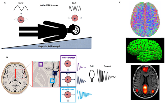
The B0 field not only affects the level of hydrogen protons, but also affects the rotation speed of these protons (this is called the common frequency rotation rate). The common frequency depends on the strength of the magnetic field. The stronger the magnetic field, the faster the protons rotate. These two phenomena are incredibly important when using MRI signals of these hydrogen molecules with high accuracy.
How do we measure the magnetic field?
So how do we measure the fine magnetic field caused by additional hydrogen protons (above) in our bodies from the massive field of B0 scanners? To do this, something called radio frequency (RF) is used. Radio frequency pulses are used to stimulate resonant protons away from the B0 field. By creating a disturbance or flip on all protons, this radio pulse causes the protons to move slightly from the orbit of the main field of B0 (fig. C). The frequency of RF pulses should be equal to the frequency of hydrogen protons spinning, so they can exchange energy called resonance or resonance frequency in physics where most energy exchange occurs. The resonance enables protons to absorb enough energy from the RF pulse to rotate their axes from the B0 field, so that the MRI scanner is able to measure it. If we think again about the compass in the Earth’s magnetic field heading toward the North Pole, if we put a small rod magnet next to the compass, we can rotate the needle to turn east. This is similar to the way protons behave when they turn on the RF pulse.
All in all, we can say that the body is full of hydrogen protons (top) that all rotate at the same dominant frequency in B0, how do we just target those in the brain to have a distinct brain image? We take advantage of the fact that the common frequency of protons is entirely dependent on the magnetic field. If a second magnetic field called B1 is applied to a certain point, hydrogen protons in that part rotate faster like the head in the chest, stomach and legs. Then, by applying RF pulse, the dominant frequency of hydrogen protons in the head can obtain a separate signal from the desired organ. Then the RF pulse will only be intensified by protons in the brain. Therefore, only protons in the brain absorb the energy from the RF pulse and move away from the B0 field. If we want to capture the legs, we can similarly adjust our RF pulse to intensify in other parts of the body, such as the legs, with proton radiation.
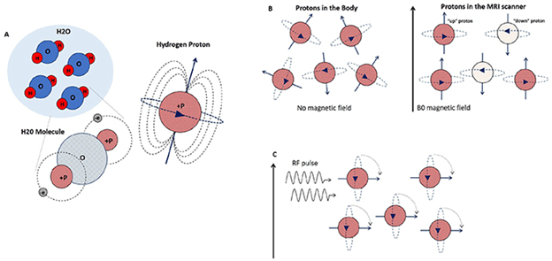
The different parts of the figure above are as follows.
(A water is composed of two hydrogen atoms and one oxygen atom. The hydrogen nucleus (shown in red as P) contains a positive charge – a proton that rotates on its axis, acting like a small magnet.
B) In mri machine, protons are aligned with magnetic field B0, some “high” (red) and slightly “low” (white). The total magnetic field derived from all hydrogen protons almost cancels each other out so that only the magnetic field remains from a small part of the additional protons (above), and this is the small magnetic field that we can measure using an MRI.
(C When an RF wave/pulse or the same proton resonance frequency is twisted, the side protons (above) move away from field B0 because they absorb RF energy.
Main question: How does the body tissue image come from these events?
All the materials expressed so far were physical laws that were simply expressed and there is almost no ambiguity for anyone. Now we want to know how these rules of seemingly simple physics are used in MRI imaging. What we do know is that we need to use protons that are in an escalation mode. When the RF pulse is off, the protons return to the main magnetic field, B0, and reshape. If we think again about the example of the compass, when we move our little rod magnet away from the compass, the compass needle rotates from east to north and once again aligns with the Earth’s magnetic field. In MRI, RF pulse has the rule of rod magnet that intensifies protons and removes their axis from the main field. When the RF pulse is cut, these protons return to their original place like our compass. This return of protons also brings energy release. This emitted energy becomes the main basis for the production of images of body tissue. That is, it is enough to use a special mechanism to absorb and image this energy. The interesting thing is that different tissues of the body lose different amounts of energy. As a result, the image of each part of the body is completely separate compared to other parts. To measure this emitted energy, we need special equipment (called coils) placed around the body part we are shooting. Inside these coils, the coils act as receiver antennas and detect the released energy as a very weak electrical current. Electrical signals are converted from computers to raw data for illustration using a complex mathematical computational set based on Fourier transforms into raw materials called Raw Data. Finally, since protons are found in a variety of tissues in the body such as gray matter, adipose tissue, hard bone tissue, etc. All different amounts of energy, the result of the converted energy is a completely accurate picture of the tissue inside the body.
غرب تهران یکی از پرتردد ترین و پرجمعیت ترین نواحی تهران محسوب می شود که به دلیل همجواری با شهر کرج همه روزه تعداد زیادی برای تردد به تهران در این مناطق در رفت و آمدند. به دلیل وسعت و جمعیت زیاد غرب تهران یافتن یک ام ار ای خوب در غرب تهران یک چالش […]
Usually, everyone in life deals with himself or an acquaintance at least once to the MRI. Now if you are a resident of Kerman city and you plan to take the turn of M.R.A. Kerman, then it is better to read this article. MRI is one of the most widely used diagnostic methods for medical […]
M.R.A. Qom If you are a resident of Qom city and you plan to take the M.R.A. of Qom, then it is better to read this article. MRI is one of the most widely used diagnostic methods for medical imaging that can accurately show many problems and diseases inside your body. Mri is a medical […]
What is the difference between CT scan and MRI? Today, there are few who can say that he has never heard of an MRI or a CT scan. We’ve all heard of these two body imaging methods. But certainly many people don’t know the difference between these two methods and sometimes mistakenly move their scan […]


