
دسته: Specialized Pet
فیلتر محصولات
در حال نمایش یک نتیجه
What is a specialized pet?
PET scan imaging is often found alongside a well-known CT or MRI machine. In this article، we will study specialized PET devices such as Breast PET، Brain PET and other specialized PET for imaging organs such as the head and neck and breast. The reason for using a CT or MRI machine in addition to a PET scan or SPECT is to locate the location of the lump or lesion found in PET scan or SPECT images.
PET scanning and SPECT devices due to the large field of view (FOV) as well as the type of images taken in nuclear medicine method, are not able to accurately locate the location of the mass or lesion. Therefore, by using CT scan or MRI, this problem can be identified by combining PET scans and CT or MRI to identify the exact location and type of the disease.
According to the above description, specialized pet devices can be divided into two groups of specialized breast pets and specialized head and neck pets. We will examine and study each one in full.
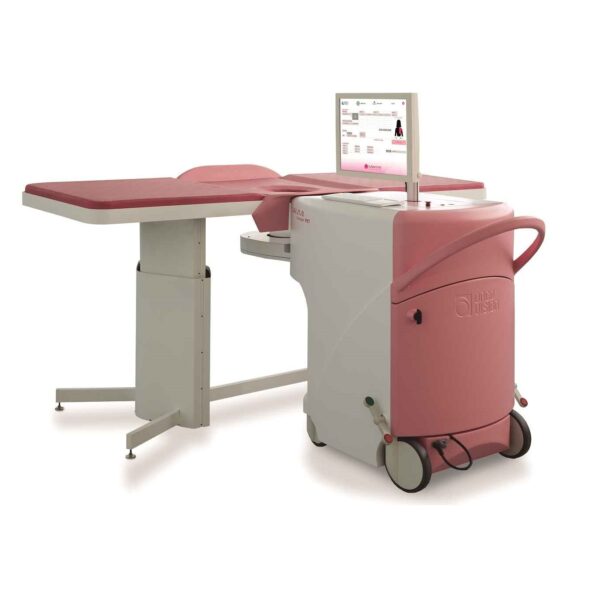
Pet Breast Specialty:
Considering the prevalence of breast cancer among women in the world and especially its prevalence at an early age, diagnosis and treatment of this problem has recently been the focus of the respected experts in the field of oncology and global health.
Considering that the treatment of this cancer is not easily possible and generally the methods used, cause the next problems for patients, and also considering the fear of recurrence of the disease that will accompany the patient throughout life, early detection of it seems the only definitive way to cure the above problem.
At the present time, in order to diagnose the above-mentioned complication by non-invasive method, two types of so-called “anatomy” and “physiological” imaging systems are used, each of which for the reasons mentioned below, lack the desired efficiency and sometimes even their combination is used to cover up the weaknesses of each other, which is not always effective.
Weaknesses of existing anatomical imaging systems:
The inefficiency of anatomical imaging systems such as MRI, mammography and ultrasound (elastography) are commonly used in this regard is due to the fact that these systems use anatomical differentiation between healthy and cancerous tissues to find them, whereas early stage breast tumors are not only very small but may not even be anatomically different from adjacent tissues. Therefore, not only are anatomical imaging systems with low resolution trapped in the detection of such small masses but perhaps because of the anatomic integrity of tissues, they do not have a proper mechanism for their diagnosis, and usually these systems are able to detect such masses when the mass volume is very large and there are more limited solutions available to treat it.
Weaknesses of existing physiological imaging systems:
In order to diagnose tumors that are very similar to adjacent tissues, physiological detection systems are used in which physiological differentiation is used instead of anatomical differentiation of tumor tissue and adjacent tissues. Of course, as previously mentioned, such devices also have limitations and defects that prevent them from being pervasive.
Among these, it can be noted that BSGI (Breast Specific Gamma Imaging) has a low resolution in which the pressure of breast tissue during imaging not only annoys the patient, but also because of the compression of the cancerous tissues, it is impossible to determine the location and shape of the tissue.
MRS (Magnetic Resonance Spectroscopy) system is another modality that is currently considered as one of the most reliable diagnostic references, but its low sensitivity to PET device, the time consuming of the imaging process, about 40 to 45 minutes under standard conditions and the positive false results that it sometimes creates, make it unreliable and ultimately cannot be easily cited.
The next modality used in the physiological diagnosis process is General PET, which has the ability to detect tumors in question, but its resolution is still in comparison to the Pet Bbw Breast Breast PET was lower (minimum 4 mm) and more importantly, due to lower sensitivity, the dose received by the patient was significantly lower than Pet Bbw Breast Breast PET (PEM) is higher. Also, due to the possibility of further injury to the patient, unlike PEM, it is not generally usable in the early stages of screening.
It seems that at present a system that can solve the shortcomings of previous systems, can achieve a high degree of confidence in the diagnosis of breast cancer in the early stages, monitor the patient until the end of the treatment process and do not harm the patient, the system Pet Breast Breast PET is designed specifically for breast imaging with the aim of reaching the highest sensitivity and resolution as two basic factors of mass detection, and in particular, the anatomical status of the desired tissue in the patient’s body and comfort during imaging are considered.
The array of detectors in it is a ring that surrounds the chest in a hole-like space and creates extraordinary resolution and high speed of imaging due to the proximity of the detectors and breast tissue.