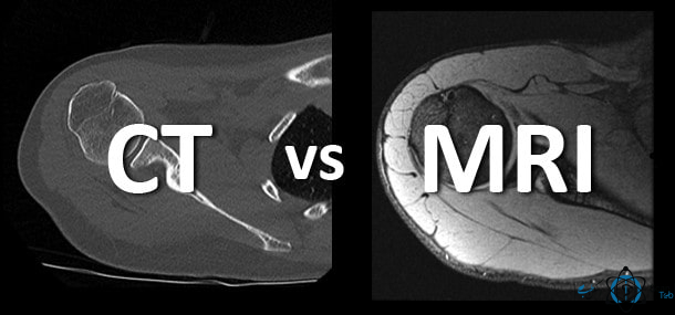
CT Scan and its introduction:
Computed Tomography scan is one of the medical imaging modalities used to create cross-sectional or shear images of the body by computer using X-ray rotation devices.
The CT device works by sending several X-ray beams at different angles, so that by irradiating these rays into the patient’s body, a 3D image is formed by a computer.
Images provide more accurate information than normal X-ray images, they can show soft tissues, blood vessels and bones in different parts of the body.
In fact, computerized tomography scans or CT scans combine a series of X-ray images taken from different angles around a person’s body and use a computer to create cross-sectional or shear images of bones, blood vessels and soft tissues such as lungs and brains in the body.
The technology of CT imaging device in continuation of radiology pathway caused the medical and treatment pathways to change dramatically. Using this method, doctors were able to easily see diseases that were often seen in surgery or autopsies in the past and to treat and diagnose more easily and quickly.
The increasing progress of CT scanners, with the passage of different generations of these devices, has been such that they can provide images with cuts less than one millimeter away from the human body.
In general, CT scan is performed in two ways without injection and by injection of contrast agent, according to the doctor’s diagnosis, a special substance known as contrast agent may be given to the patient in order to detect different parts of the body more clearly in X-ray images.
Due to its nature, contrast agent blocks X-rays and causes its image to be seen white on CT images, making the organ given to it completely recognizable from other organs, such as the intestines, blood vessels or other organs examined.
Depending on the part of the body that is examined, the patient may need to drink a liquid containing contrast agent. It may also be necessary to administer contrast agent by injection into the arm or anus through the right intestine. If your doctor intends to use contrast agent, he or she may ask you to take a 9-6 hour before a CT scan.
Why is a CT scan done? Applications of CT scan:
Currently, more than 75 million CT scans are performed annually in the United States. According to one of the most reputable American journals, 2% of cancers (malignant type) are due to high doses used in CT scans. The combination of PET and CT, known as PET/CT, is a new method in which anatomical information that can be detected from CT images is combined with metabolic information (using pet scan images) related to function and components and is a desirable method, especially in the study of tumors. It is important to note that high dose of CT is not necessary in this method. This method is used 85% in the study of tumors (diagnosis of malignant cells from benign, accurate observation of the progression of cancer cells and evaluation of response to treatment and radiotherapy), 10% in neurology, 5% in heart studies.
CT scan has many applications, especially for diagnosis of diseases and evaluation of injuries is very suitable. This medical imaging method can help your doctor to:
Diagnoses infections, muscle disorders and bone fractures
Determine the location of masses and tumors (including cancer)
Study blood vessels and other internal structures
Assess the extent of internal damage and internal bleeding
Guide surgeries and biopsies
Monitor the effectiveness of treatments for some medical conditions, including cancer and heart disease, pulmonary nodules and liver mass.
Medical imaging with CT scan is almost noninvasive and can be done quickly.
How is a CT scan done?
Modality CT is one of the medical imaging modalities that is used to create cross-sectional or shear images of the body by computer using X-ray rotation devices.
CT scan works by sending several X-ray beams at different angles, so that by irradiating these rays to the patient’s body a 3D image is formed by a computer.
Images provide more accurate information than normal X-ray images, they can show soft tissues, blood vessels and bones in different parts of the body.
In fact, computerized tomography scans or CT scans combine a series of X-ray images taken from different angles around a person’s body and use a computer to create cross-sectional or shear images of bones, blood vessels and soft tissues such as lungs and brains in the body.
CT scan technology in continuation of radiology pathway caused the medical and treatment pathways to change dramatically. Using this method, doctors were able to easily see diseases that were often seen in surgery or autopsies in the past and to treat and diagnose more easily and quickly.
More information about this CT scan device can be found here .
What is the difference between CT scan and MRI? Today, there are few who can say that he has never heard of an MRI or a CT scan. We’ve all heard of these two body imaging methods. But certainly many people don’t know the difference between these two methods and sometimes mistakenly move their scan […]