All about Pet Scan
PET scan is a nuclear medicine imaging technique that provides a three-dimensional picture of the function of the organs of the body. The system detects a pair of gamma rays produced by a radionuclide of positron ten (tracer) and emitted in opposite directions.
This radionuclide is inserted into the body by an active biological molecule. Then, the 3D image of the drug accumulation inside the body is reconstructed by computer analysis. In newer scanners, a CT scan is usually taken simultaneously in the same device to make the 3D image more accurate.

PET (Positron Emission Tomography) is one of the most advanced imaging systems that has demonstrated unique capabilities in the diagnosis of cancer, neurology and cardiovascular diseases. More than 2,700 PET centers have been established in the world that provide clinical services.
Most PET centers are active in the United States, Germany and Japan, and in the region of Saudi Arabia, the United Arab Emirates, Qatar, Bahrain and more recently in Egypt.
What is PET scanning?
Positron emission tomography (PET) scans or PET scans produce detailed 3D images of the inside of the body.
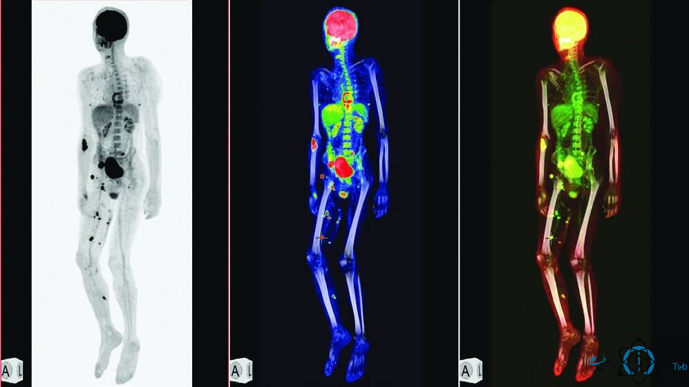
PET scans can clearly show the part of the body being examined, including any abnormal areas, and can identify specific functions of the body.
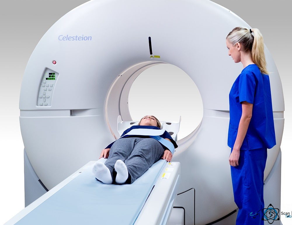
PET scans are often combined with CT scans to produce more detailed images. A PET scan is also known as a PET CT scan.
PET may also sometimes be combined with an MRI machine، which is called the final device of the PET-MRI.
Principles of PET System Operation
Radiotherapy to the patient:
- In PET scan imaging, the patient’s body is injected with a special chemical labeled with a radioactive substance (radiopharmaceutical). Different tissues of the body absorb different amounts of this substance due to their blood flow rate and their cellular and chemical metabolism. The absorbed material emits an invisible beam that can be received by a special imaging device.
According to this Introduction, the principles of PET imaging can be based on the detection of invisible beams resulting from the destruction of positron and electron pairs, hence the radioisotopes used in PET are C11, N13, O15 and F18 which have half-lives of 20, 10, 2 and 110 minutes, respectively.
What is the function of radiotherapy in the patient’s body?
The radioactive elements are not chemically different from the non-radioactive type and are naturally present in the human body, and even the air we inhale is a mixture of carbon, nitrogen and oxygen. Some of these radioisotopes are used directly or by adherence to drugs or substances that are absorbed into a particular organ or area of the body or are used to check for specific conditions of defects.
After the radiopharmaceuticals were injected or gassed through respiratory system to the patient, this radiomedicine is absorbed into the organ or the imaging site and is distributed and absorbed according to the conditions of the disease in the studied organ.
Radiopharmaceuticals injected in PET scan emit positrons and positrons are paired with electrons and destroyed, resulting in the destruction of each pair of positron and high-energy electron (511 KeV) in the opposite direction.
Therefore, a large number of dependent pairs of beams are emitted in different directions from the patient’s body. If there are detectors around the patient, by revealing these beams, it is possible to obtain a three-dimensional image of the distribution of radiopharmaceuticals in the examined organ by which the presence of defects or disease can be detected.
If the biologically active molecule for PET imaging is F18 (FDG) (of glucose compounds), the image shows the metabolic activity of the tissue in areas where glucose uptake is high. This substance is a labeled derivative of glucose which is very low and in general and normal conditions there is no significant risk in using this technology.
The most common type of PET scan is the use of FDG to look for cancer metastasis, accounting for about 90% of current scans in standard medical care. However, in other cases, other markers, due to the accumulation properties at a particular point, can be used to capture images of points within the body.

PET Imaging Compared to Other Medical Imaging Modalities
In most diseases and disorders, changes in metabolism occur before anatomical changes occur in the tissue, so unlike conventional imaging systems such as MRI and CT, anatomical changes or SPECT measure physiological changes.
With the PET system, we will be able to detect the disease at an early stage, prevent and treat the progression of the disease, and as we know it, early detection is critical for treatment.
Comparison of PET Imaging with SPECT:
If we compare the PET system with SPECT, which is currently the most common system in nuclear medicine, the following should be considered:
1- The spatial resolution of the PET system, which is the ability of the system to show the details of the studied organ in the image, is about 4 to 5 mm, which is twice as good as the spatial resolution of SPECT.
2- PET efficiency is far better than SPECT system which results in greater resolution and contrast of images.
3. Due to the possibility of correction for absorbing radiation in PET scan, quantitative analysis of information is possible which has many applications in determining the stage of the disease.
In addition to studying the physiological process of the tissue or organ studied, there is the possibility of studying the metabolism and biochemical activity of the studied tissue, which is the most prominent advantage of this system which distinguishes it from other imaging systems.
5. Because the injected radioactive material has a very short half-life in the PET method, the shelf life of the substance itself and its radiation in the body will be low. However, in order to remove the effects of radioactive substances faster, the patient will need to drink plenty of fluids after the test.
What are PET Scan Applications?
PET scan is a non-invasive imaging technique that is widely used in the diagnosis, treatment and follow-up of multiple diseases in clinical oncology (medical imaging of tumors and the search for metastases) and clinical research of brain diseases such as Alzheimer’s and Parkinson’s, as well as in cancerous, cardiovascular and neurological diseases.
PET is also used in pre-clinical animal research, where it is possible to repeat experiments on a particular subject. This is especially valuable in cancer research because the statistical quality of information (subject matter is used as self-control) and consequently the number of animals required for a particular study decreases.
Some imaging scans, such as CT and MRI, show anatomical changes in tissues within the body, while PET and SPECT are able to detect biological molecular changes (even before anatomical changes). PET scans do this with radioactive labeled probes whose absorption rates vary across different tissues.
In a PET scan, changes in regional blood flow in different anatomical structures (as a measure of the injected positron emitter) are observed and quantified. PET images can be obtained with a biceps gamma cam fitted with a synchronous collision detector, but the image quality is considerably lower and the time to obtain more image. This is a good option for institutions that have less demand for PET rather than referral patients elsewhere.
Some common applications of PET include oncology, neurology, cardiology, pharmacology, imaging of small animals, musculoskeletal imaging and cancer diseases, especially gastrointestinal, breast and lymphoma cancers.
What types of tumors does the PET scan examine?
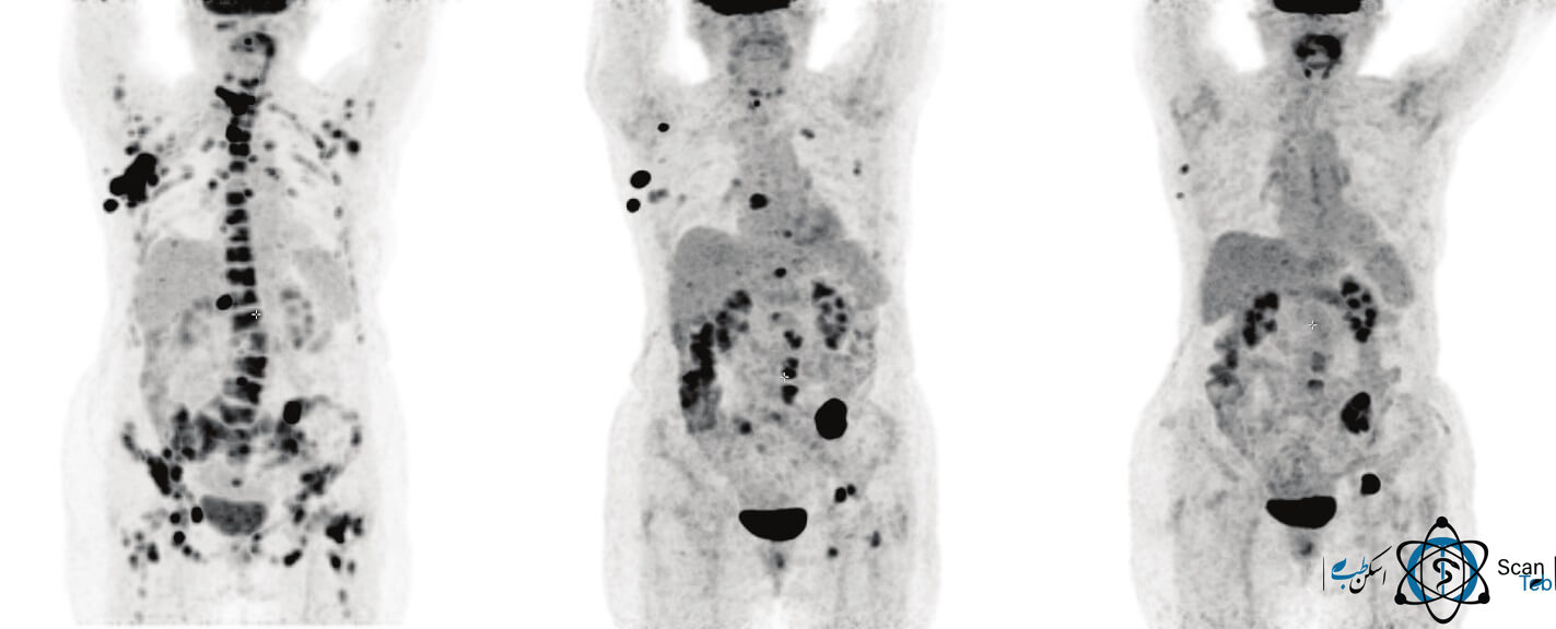
The result and report of the PET scan image can help diagnose cancer and its spread rate. PET scans can reveal tumors in the brain, prostate, thyroid, lungs and cervix. The scan can also assess the occurrence of colorectal, lymphoma, melanoma and pancreatic tumors.

PET Scan
How long does a PET scan take to diagnose cancer?
How long does a PET scan take? This is one of the most important questions for patients and patients’ companions who come for the first time for PET scans. Depending on the type of information your doctor is looking for, a PET scan can take about 60 to 90 minutes.
After the radiotherapy is injected, you will need to wait about an hour in the rest room after the injection, until the radiotherapy is spread throughout the body and you are ready to be scanned. After this time, the device expert will ask you to go to the scan room for PET scanning. Once you’re ready to start the scan, you’ll be asked to lie on your back on the bed of the PET CT machine and sit still until your scan is complete.
Is a PET scan painful?
Generally PET scans are not painful, but depending on the patient’s condition it may be frustrating. PET scans are a type of nuclear medicine scan، as we have discussed in the articles presented in the Medical Scan، with F-18 or C-11 radiopharmaceuticals or various other radiopharmaceuticals. PET scans or PET scans are not harmful.
But in some situations they can be uncomfortable or boring. For example, for a full-body or whole-body PET scan, the patient must lie still during the scan. You may also need to keep your arms above your head. These may cause fatigue and, in some cases, pain during the scan.
Security in PET scan imaging:
PET is a non-invasive and safe procedure, even in the use of radiopharmaceuticals related to it, there have been no severe adverse reactions and adverse reactions. This is because they are usually used in micrograms. Some studies have noted a very low risk of developing secondary cancer caused by radiopharmaceuticals, which has not been conclusively proven (Facey K et al 2007).
Although this technology is generally safe and risk-free, it is necessary to use the following precautions (Facey K et al 2007):
- Not used in pregnancy.
- After the test, the patient should stay away from the children for several hours.
- Careful use of FDG in patients with diabetes intolerance.
- Personnel working with a PET machine should be given the necessary training on the preparation and use of radiopharmaceuticals and how to transport them.
What are the limitations of PET scanning?
The limitations of the use of PET scans are mostly related to the high cost and high price of cyclotrons, which are responsible for producing radionuclide with a short half-life. There is also the cost of chemical synthesis in place for radiopharmaceuticals (tracer) after radioisotope production. Organic radiotracer molecules should not contain the radioactive isotope from the beginning, because then its bombardment by cyclotron would destroy its organic careers.
Instead, the isotope must be produced first (the bombardment of the radioactive nucleus by the cyclotron) and then the chemistry lab synthesizes the necessary tracer (e.g. FDG) from it shortly before the radionuclide decays. Few hospitals and universities can afford the cost of cyclotron maintenance, and most clinical PETs are supported by third-party radiopharmaceutical manufacturing institutions that can provide services to multiple centers at the same time.
These limits limit the use of PET to fluorine-18, which has a half-life of 110 minutes and has reasonable mobility before use. Rubidium-82, which is produced in portable generators and used in Myocardial Perfusion can also be used.
How much does a PET scan cost?
In general, the cost of PET scanning is expensive around the world, and one of the reasons for the delay in the arrival of these equipment to Iran is the high cost of equipment (PET CT and cyclotron and its accessories) and the high cost of these tests.
On average, scanning costs around the world and countries such as the United States, Turkey, Japan, etc., PET CT scanning can cost between $2,000-$5,000 or more. This includes the cost of medicine and scanning.
The cost of PET scanning in Iran is not covered by insurance, depending on the type of private and public centers in Iran and the type of radiopharmaceuticals in these centers.
Approximate cost of scanning in PET scan centers in Iran is as follows:
The cost of PET scanning with radiopharmaceuticals in Imam Khomeini Hospital and Shariati Hospital is about five million tomans.
The cost of PET scanning with radiopharmaceuticals at Khatam Alanbia Hospital in Tehran is about 9.5 million Tomans.
The cost of PET scanning with radiopharmaceuticals in the Karaj Payam Center is about 7.5 million Tomans.
The cost of PET scans at Masih Daneshvari Hospital varies depending on the type of insurance, but it is free of charge of five million and two hundred and fifty thousand tomans.
In general, the cost of PET scan according to the tariff approved by the Ministry of Health for government centers varies depending on the type of radiopharmaceuticals used. The Scan Teb team has provided you with the cost of PET scan with different drugs and a 1400 tariff.
The cost of PET CT with FDG is 52.000.000 Rials.
The cost of PET scan with Galium PSMA and DOTATATE is 67.500.000 Rials.
Brain and whole body scans are 80,770,000 Rials.
The cost of PET scans for foreign patients varies in different centers, which you can contact the support team of this platform for this amount. You can also choose your preferred center to receive a PET scan via the link to receive a PET scan.
How long does it take to get the PET scan results؟
When should I get my PET scan results? This is the first question you may ask to accept the PET scan after undergoing a PET scan. Usually, depending on the type of PET scan centers and crowds, this question will have a different answer. The best and most accurate answer to this question is the answer that the receptionist will give you.
But according to medical scan studies, nuclear medicine specialists with the help of radiologists will respond to images processed by PET CT processing systems, usually within 24 to 72 hours after the scan, and you can get your PET scan report within this time period from the center or reception or the system that the medicine scan provides to the centers.
For more information about PET scans and scientific study in this area, suggest scanning medicine study website Mayoclinic .


