What is 4D ultrasound and what is its application?
What is 4D ultrasound and what is its application? This is a question that expectant mothers and even their wives may ask themselves. No doubt if you’re a pregnant lady, you’re impatient to see your baby growing in your belly – by the way, nine months is a long time to wait for a little glimpse of those lovely fingers and feet of your child.

Since 3D and 4D ultrasound scans allow you to see your unborn baby in more depth and detail than a two-dimensional ultrasound, you may be keen to have a photo of your unborn child on the album.
But before doing so, it’s important to know when to use 3D ultrasound and 4D ultrasound during pregnancy to ensure that you do everything you can to keep yourself and your baby safe and sound.
What is the difference between 2D, Doppler, 3D and 4D ultrasound?
During pregnancy, you may experience a combination of the following ultrasounds:
2D Ultrasound:
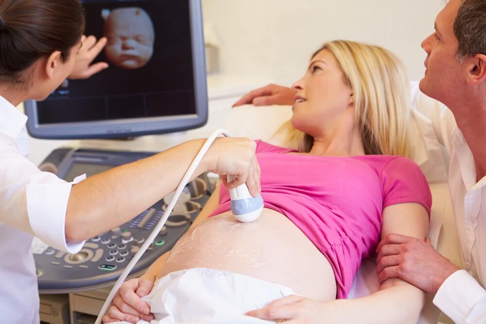
If you’ve visited a doctor, you’ve probably already experienced a normal (2D) ultrasound and you know it can be an exciting and magical moment. For this scan, a probe (transducer) is placed on your abdomen or inside the vagina to send sound waves in your body. Waves are reflected from internal organs and fluids, and a computer converts these echoes into a two-dimensional image (or cross-sectional view) of the fetus on the monitor.
Doppler Ultrasound: With Doppler’s fetal ultrasound, your doctor will use a handheld ultrasound device to amplify the fetal heart rate with a special gel on your abdomen.
3D Ultrasound:
For 3D ultrasound, several 2D images are taken at different angles and then combined to form a 3D rendering. In this article posted on the website of Scan Teb, you can learn in detail about 3D ultrasound of the fetus.
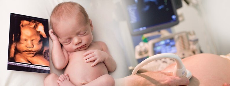
4D Ultrasound: 4D ultrasound is similar to 3D ultrasound, but has a capability beyond and can show motion picture like a video. So in a 4D ultrasound, you’ll see your baby doing things in real time (like opening and closing his eyes and sucking his thumb).
Reasons for ultrasound during pregnancy
In general, women with low-risk and uncomplicated pregnancies usually perform ultrasounds at least once, while older mothers and those with complications usually perform more ultrasounds. There are many reasons why ultrasound is generally necessary during pregnancy, depending on the length of pregnancy, including:
- Predicting the date of delivery
- Listen to the heartbeat
- Ensure that the pregnancy is not outside the uterus (i.e. in the fallopian tubes) and in the uterus.
- Confirmation of the number of embryos in the uterus
- Ensure proper embryonic growth rate
- Evaluation and measurement of the main organs of the newborn
- Measuring baby size
- Evaluation of amniotic fluid levels
- Rejection of some congenital defects
- Determining the gender of the baby
Why is 3D and 4D ultrasound performed during pregnancy?
Physicians use 2D and Doppler ultrasounds in uncomplicated pregnancies to examine the fetus, evaluate amniotic fluid and assess congenital defects and other reasons.
3D and 4D ultrasounds are performed only to closely examine suspected fetal abnormalities such as cleft lip and spinal cord problems, or to monitor specific cases. In other words, 3D ultrasound and 4D ultrasound are usually not part of routine prenatal examinations.
Is 3D and 4D ultrasound safe during pregnancy?
Depending on what we know, experts say that ultrasound should only be performed by a qualified medical professional that your doctor will recognize as essential for medical reasons. Currently, doctors recommend that women expect at least a two-dimensional ultrasound between the 18th and 22th weeks of pregnancy, given that some women may also undergo a first-trimester ultrasound.
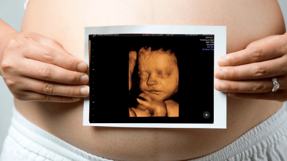
In general, 4D ultrasound, such as 3D ultrasound, is safe. But keep in mind that an ultrasound can’t be a good reason just because you take a photo of the fetus. You should have a good medical reason for a 4D ultrasound. But if you have a 4D ultrasound, you can keep the fetal film inside the mother’s abdomen as a memento for years to come.
Final word:
The medical scan team hopes to take full advantage of this article and at the end of the article it is essential to point out that the scan of medicine by providing the medical imaging turn-by-turn platform for patients and centers active in the field of medical imaging in this area has tried to educate the patients who intend to take the ultrasound turn and its types, mri turn and its types such as MRI for high weight patients and CT scans and things like that have to help them. With the support of our strong scientific team across the country, we have tried to introduce medical scan centers and types of scans.
You can use
the platform to get a scan
of different types of 3D, multidimensional and different types of ultrasound, MRI, CT scan, nuclear medicine and pet scan, radiology and angiography in your city. We hope that everyone can choose the best and nearest center to themselves in their city and get their scan turn without any hassle.