Kemer MRI + Interpretation of Kemer MRI Response
With the increasing availability and use of MRI imaging, physicians today use back MRI to diagnose the causes of back pain. In this article, we will try to familiarize you, however briefly but specializedly, with the interpretation of the MRI answer and show you the specialized terms and images about the back and spine MRI.
MRI may be useful in certain clinical situations in low back pain, however, the importance of a thorough clinical evaluation cannot be done with MRI alone. Understanding the benefits and limitations of MRI in assessing low back pain and improving communication among physicians should provide optimal management of clinical problems similar to patient’s radiology.
Several methods are used for spinal imaging, and with increased access to MRI imaging and better imaging quality, doctors commonly use MRI to find the roots of spinal and back pains.
Patients with spinal injuries often visit a doctor with a range of symptoms. Differentiation of the three common symptoms of low back pain, sciatica and limping is very useful for physicians as they can help determine the origin and cause of the patient’s symptoms. While MRI is accurate in diagnosing spinal pathology, sometimes the MRI image of the spine discovers other conditions that may not have a significant clinical impact and sometimes confuse doctors.
Low back pain can be caused by several diseases, ranging from arthropathy of the facet joint to muscle strain. Pain is mainly as the term suggests in the back and back and comes from locally influenced structures. On the other hand, sciatica has a different pattern of pain in terms of distribution and is caused by nerve root stimulation. This can occur due to the direct compressive effects of intervertebral disc herniation on the nerve root or an underlying inflammatory process, such as an infection that causes acute pain in dermatoma distribution.
Limping or limping is traditionally divided into two categories: Neurogenic or vasogenic, which depends on the underlying cause. Limping is often described as impaired mobility and vague pain in the lower extremities. Central vertebral canal stenification is a common cause of neurogenic lameness and has a different pattern, while vascular lameness is more consistent and repeatable.
The importance of determining the chronic symptoms and identifying them in history and clinical examination, such as fever and paresthesia perineum, is very important in formulating clinical diagnosis and differentiating benign causes such as musculoskeletal pressure. For example, epidural abscesses or spinal metastasis can cause chronic back pain. Some risk-enhancing factors such as the patient’s age and history of medication (e.g. steroid use) may also be suspected of ankylosing spondylitis or compressive fractures. Studies show that MRI is a highly sensitive but less specific imaging method for evaluating lumbar spine conditions.
Anatomy of the lumbar spine
The lumbar spine consists of five distinct vertebrae separated by intervertebral discs and strengthened by multiple ligaments and paravertebral muscles. The bone sac containing the medularis cone and nerve roots is located in the central vertebral canal. The nerve roots are then removed from the spine through the intervertebral hole canal instead of the vertical angles seen in the cervical spine. Understanding this anatomical relationship allows the doctor to separate the nerve root stimulated by an intervertebral disc herniation.
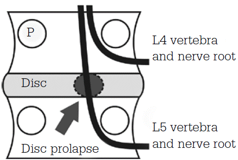
Figure 1 Relationship between The Origins of The Outlet Nerve, Pedicle (P) and Intervertebral Disc
The outgoing nerve roots pass through the nerve hole and this is divided into parts based on its association with the stem and zigapophysical joint at axial and sagittal levels (Fig. 2). In the axial plate, the outgoing nerve root trads the sub-joint trough from the central area to the perforated area and out of the hole. Subsemal, super-pedicular, pedicular and disc surfaces are used to isolate areas along the longitudinal axis.
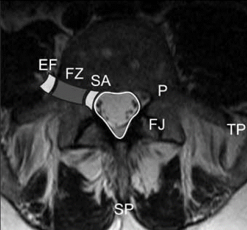
Figure 2. T2-weight axial incision from the lumbar spine that shows different regions along the pathway of the nerve roots output from the subarm joint (SA) to the perforated area (FZ) and the outer perforated area (EF).
Intervertebral discs have a hydrated pulposus nucleus located in the allied rings of the fibrous cyclic center. As we age, the discs gradually dehydrate, resulting in a decrease in the T2 signal, which is often observed in asymptomatic patients.
Disk Pathology
The nomenclature used in spinal imaging reports is sometimes confusing. For a better understanding, the following standard nomenclatures can be used:
- ‘Natural’
- Congenital/developmental type (Congenital/developmental variant)
- Degenerative – herniation, hernia, degenerative – annular fissure, herniation, degeneration)
- Inflammation/infection
- Neoplasia
- Morphological type of unknown significance (Morphological variant of unknown significance)
- Spinal annular cleft or Annular fissure
Any separation between the annular filaments or the separation of circular fibers from the vertebral body is defined as an annular incision. These changes often occur in degeneration conditions asymptomatic disks. Therefore, the term “ring rupture” does not arise due to the presence of a traumatic stimulant.
Disc herniation or disc herniation
Disc herniation usually occurs in two scenarios in which the spine is traumatized or damaged in the form of abnormal axial loading or secondary dynamic change to congenital or acquired spinal abnormality. Hernia or rupture causes nerve root compression and pain.
Any disc material that extends beyond the vertebral bodies is considered a disc herniation. This phenomenon is mostly described as “disc bulge,” “bulge” and “extrusion.” The basic purpose of using these terms is mainly a descriptive goal. The amount of disk expansion is evaluated peripherally at the edges of the end plate of the vertebrae (ring apophyse) at the beginning. The term “bulging disk” is used to describe disk expansion of about 50 to 100% loop apophyses. Displacement between 25-50% is described as “extensive hernia” and less than 25% as “focal hernia”.
A protruding disc has a wider base compared to the extent of the disc material spreading outside the vertebrae (Fig. 3). Conversely, when the disk expansion rate is greater than the base of the disk extension, it is described as “extruded”. When there is a separation between the herniated disk and the parent disk, it is described as “compact” (Fig. 3).
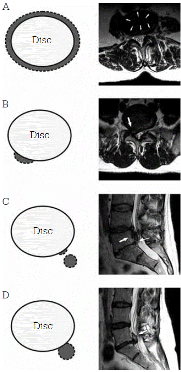
Figure 3. Axial image and MRI weighing T2 of the lumbar spine that shows disc herniation.
A = bulging disk; C = Disassembly disk material. Linear arrow: Separation between disc herniation (flash block) and intervertebral disk space. D = Extrusion
In attempts to link clinical findings and radiological evidence, the relationship between disc herniation and neural root is carefully investigated. Any contact, displacement or inflammatory changes are reported to allow accurate identification of the patient’s symptoms in the annoying pressure disc lesion.
Central vertebral canal stenity
The gradual development of central vertebral canal stenification, in which compression of nerve roots in the bone sac (Fig. 4) often leads to decreased mobility and nervous limping. In cases where an acute event or accident has occurred, the initial form may occur as horsetail syndrome, in which case immediate decompression is required through surgery.
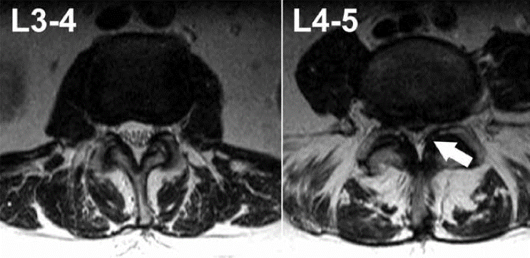
Figure 4. Axial incisions weighing T2 from the same lumbar spine of the patient at different levels. There is severe central nut canal stenosis at L4-5 (arrow) levels without cerebrospinal fluid and crowded ponytail nerve roots. In L3-4, nerve roots can be seen as low signal points surrounded by clear cerebrospinal fluid.
The causes of central vertebral canal stenification can be divided into congenital and acquired conditions such as neoplastic and degenerative changes. Degenerative changes include facet osteophytes, flavum ligament hypertrophy and disc herniation. While some diseases have a specific cause, most central vertebral canal stenosis is caused by a combination of conditions.
The intensity of central vertebral canal stenage is visually graded in the lumbar MRI image and is not currently used by the Global Rating Scale. Several centers have evaluated different grading methods, including cross-section measurement and local bag morphology. However, the use of imaging alone is inadequate to assess severity, as there is often a dissonance between mri symptoms and findings.
Ankylosing spondylitis
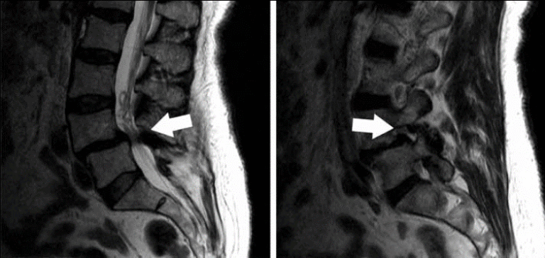
A simple radiograph of damaged joints for patients suspected of ankylosing spondylitis is often sufficient and there is no need for MRI. However, doctors mostly use um Arai to diagnose this condition and monitor the therapeutic response. The main features of lumbar spine MRI include characteristics that show underlying inflammation and its effects, such as bone marrow, squared vertebrae (Romanos lesions), sandsmophyte formation, ankylose and erosion (Fig. 5). The clinical and imaging aspects of this disease are very complex and outside the scope of this article.
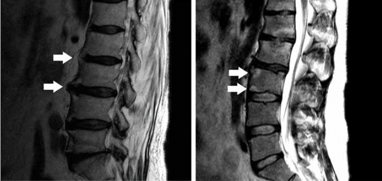
Figure 5. Sagittal T2 images of the lumbar spine in two separate patients with ankylosing spondylitis. Left: Arrows point towards poldling syndesmophytes. Right: Arrows point towards brain changes near the end plate, consistent with inflammation
Spondylolistis
Spondylolistissis is defined as a condition in which lumbar spine disharmony occurs in the form of a vertebrae that exits from its normal position relative to the lower vertebrae. This can lead to narrowing of the lateral nerve hole and central channel of the spinal cord (Fig. 6). In addition, lumbar spondylosis defects are commonly associated with spondylolistis. The chronic nature of pain experienced by the patient as well as complex mechanical issues related to spinal disharmony can often lead to failure of conservative treatment and surgical fusion of damaged surface.
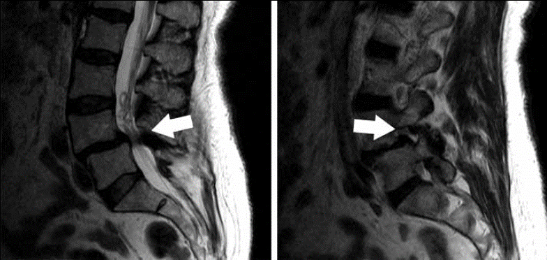
Figure 6. MRI weighing sagittal T2 from the lumbar spine. Anthrolistsis is available at L4-5 levels, leading to severe central channel stenosis and nerve holes associated with neurological collisions.
The role of MRI is to determine the severity of stenosis of the central spinal canal or nerve puncture and identify a potential cause of back pain. However, due to the constant nature of MRI imaging, some defects are not detectable during the movement of the vertebrae. Because during MRI imaging, the patient lays on the MRI bed completely and the possibility of injuries during the activity of the vertebrae cannot be done. Spinal surgeons have used dynamic X-rays or lumbar spine fluoroscopy to assess any abnormalities in the moving spine, implying greater stenification and nerve collision.
Final Word
In this paper, we tried to specializely investigate the mri of the back and the perception and sensuality of the lumbar MRI answer. But this article has covered only part of these issues. Therefore, in order to better understand the outcome of your back MRI, you need to show your back MRI image to a radiologist. If you have received a lumbar MRI scan, you will no longer need to go to the imaging center to have an MRI answer. In the scan of medicine, your MRI answer will be sent to you via SMS and you can show this SMS to your doctor so that he or she will see your MRI answer.
