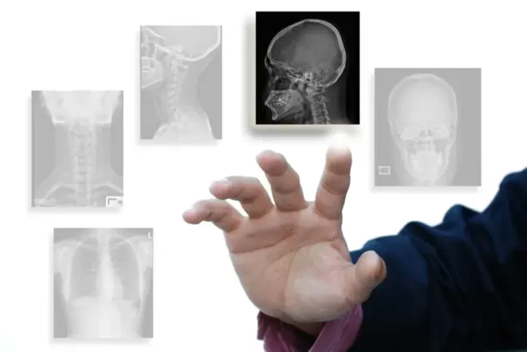
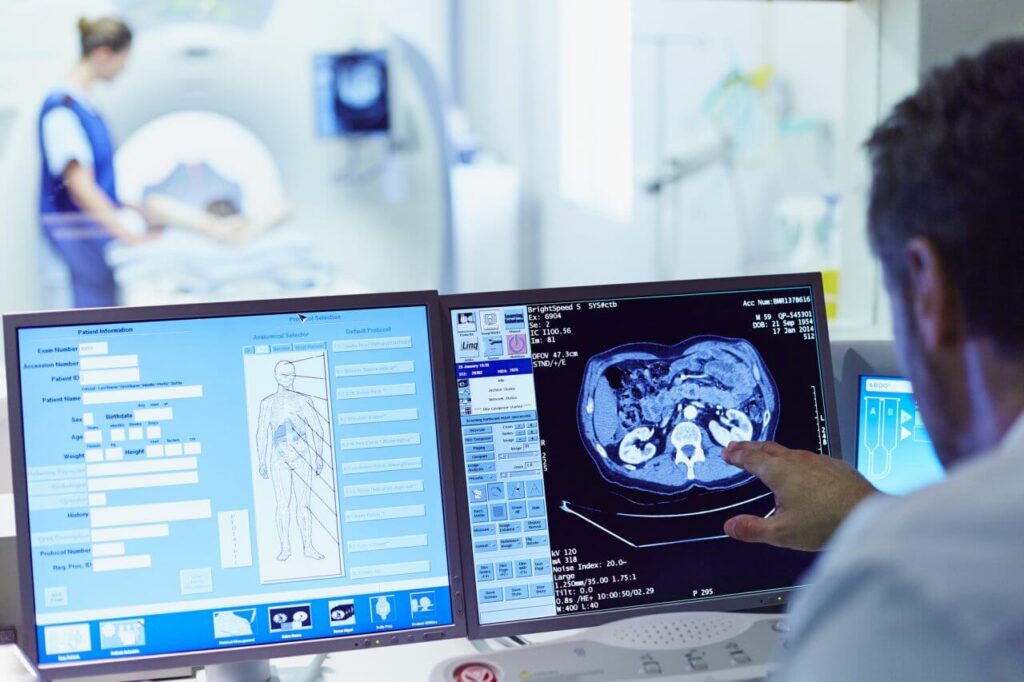
What is a bone CT scan?
Bone CT scans can provide more detailed information about bone tissue and bone structure than bone radiography, thus providing more information about bone injuries and/or diseases. But let’s explain what the difference between CT scan and radiography is.
CT scan
is a non-invasive diagnostic imaging technique that uses X-rays and computer technology to produce shear images of body tissues. The CT scan shows detailed images of different parts of the body, including bones, muscles, fats and organs. CT scans are more detailed than conventional radiology.
In conventional radiology, an energy beam shines toward the part of the body that is studied, and a plate behind the part of the body that is irradiated records changes in the energy beam after passing through the skin, bone, muscle and other tissues. While a lot of information can be obtained from a radiography, it still is not enough information.
In CT scans, the X-ray beam moves circularly around the body. This causes many different shots to be filmed from the same limb. X-ray data is sent to a computer that interprets X-ray data and displays it in two-dimensional (2D and 3D) forms on the monitor. CT scans may be performed with or without contrast.
Contrast refers to a substance that enters the body in an injectable or oral form, making the specific organ or tissue studied more clearly. Other related methods that may be used to diagnose bone problems include X-rays of bones,
MRI
imaging of bones and bone densitometration.
What are the reasons for using a bone CT scan?
CT scans of bones may be used to assess bones, soft tissues and joints in terms of injuries, lesions, fractures or other abnormalities. This scan is particularly useful when another type of scan, such as radiology or physical examination, fails to provide enough information. Also, there may be other reasons your doctor will prescribe a CT scan of bones, joints or soft tissue.
What are the risks of CT scans?
In general, it should be said that exposure to X-rays for a long time increases health risks. But these risks are not the same for everyone. If you are pregnant or suspected of pregnancy, you should inform your doctor. Exposure to radiation during pregnancy may lead to congenital defects for the fetus. If bone CT is needed, special precautions are taken to minimize the fetus’s exposure to radiation.
If contrast agents or contrast agents are used, there is a risk of an allergic reaction to these substances. Patients who are allergic to these medications should inform their doctor. If you have ever reacted to contrast agents or kidney problems, you should inform your doctor.
Patients with renal failure or other kidney problems should inform their doctor. In some cases, contrast agent can cause kidney failure. Also, patients taking diabetes metformin (glucophage) should notify their doctor before performing a bone CT scan with contrast agent, as it may cause a rare condition called metabolic acidosis. If you are taking metformin, you will be asked to stop taking it at the time of the scan and do not take it for up to 48 hours after the injection. Blood tests may be needed to check for kidney function before restarting metformin.
How to prepare for bone CT scan?
If you are pregnant or think you may be pregnant, please consult your doctor before planning a bone CT scan. Other options will be shared with you and your doctor. You may be asked to wear a patient’s gown. In this case, a special hospital gown is provided. Please leave all jewellery and precious objects at home. CT scans are often performed with and without contrast agents. Contrast agent improves the ability of the radiologist to view images inside the body.
Some patients should not have iodine-based contrast agents. If you have problems with your kidney function, please notify the imaging center representative when you make an appointment. You may be able to perform the scan without contrast agents or do an alternative imaging test.
If you have reacted strongly or anaphylactically to any contrast agent in the past, bone CT scan contrast will not be performed by injection. If you have had mild to moderate reactions in the past, you should probably take the drug before a CT scan. If your doctor has ordered a CT scan without contrast, you can eat, drink and take your prescribed medications before the examination. If your doctor has prescribed a contrast CT scan, do not eat three hours before the CT scan.
Diabetics should eat breakfast or light lunch three hours before the scan time. Depending on oral diabetes medications, you may be asked to stop taking the medication up to 48 hours after a CT scan. All other patients can take their prescribed medications as usual.
There may be other risks depending on your specific medical condition. Before performing the scan, make sure to share any concerns with your doctor.
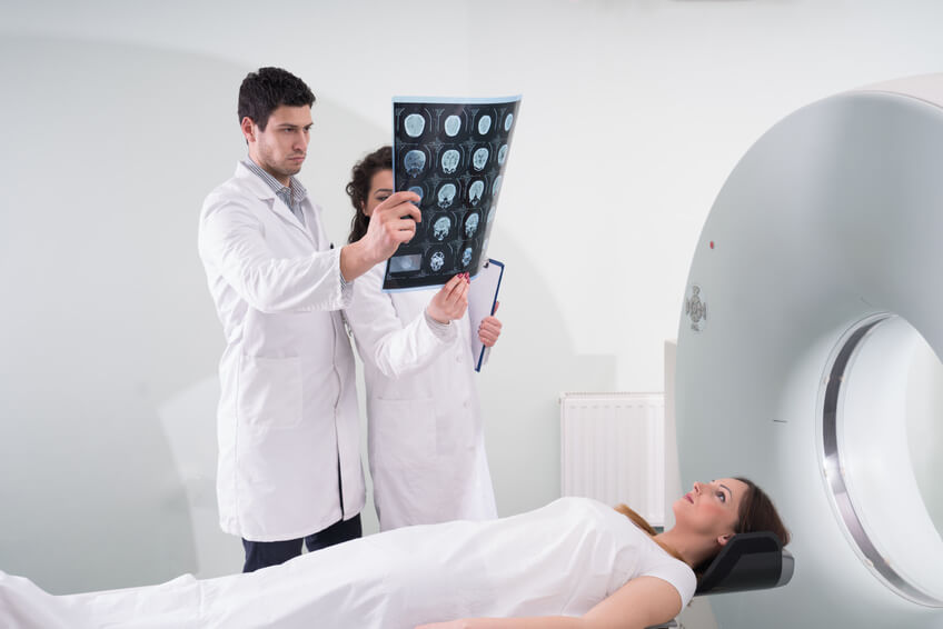
What happens during bone CT scans?
Bone CT scans may be performed outpatiently or as part of your hospitalization. Procedures may vary depending on your circumstances and your doctor’s performance.
In general, a bone CT scan follows this process:
You lie on a bed of a scanning device that goes into the large, circular span of a CT scan machine (Gantry). Pillows and straps may be used to prevent movement during operation.
The CT scan expert is placed in another room and in this room is in full control of the CT scan device. However, you will be constantly exposed to the CT scan expert through a window. Speakers inside the CT scanner allow the technician to communicate with you and hear your voice. You may have a call button so that you can notify the technician in case of any problems during the scan. The technician will always monitor you and will be in constant communication.
When the device starts to rotate around you, the X-ray will pass through your body for a short time. You’ll hear very few sounds that are normal.
X-rays absorbed by body tissues are detected by CT scan detectors and transmitted to computers. The computer converts information into an image to be interpreted by a radiologist.
It is important to remain very sedentary during bone scans. You may be asked to hold your breath at different times during the operation.
If contrast agent is used to scan your bone, you may feel some effects when injecting contrast agent. These effects include feelings of hot flashes, salty or metallic taste in the mouth, brief headaches, or nausea or vomiting. These effects usually last for a few moments.
If you feel any respiratory problems, sweating, numbness or palpitations, you should notify the technician immediately. After you’ve finished the process, you’ll be removed from the device.
While the CT procedure itself does not cause any pain, the need to lie down for a long time may cause discomfort or pain, especially in patients with bone fractures or back pain. The CT scan expert usually uses all possible convenience measures and completes the procedure as quickly as possible to minimize any discomfort or pain.
What happens after a bone CT scan?
If contrast agents are used in bone CT scans, you may experience headaches, skin rashes or other mild effects for a short time after the scan. If you see any pain, redness or swelling at the injection site after returning home after a bone CT scan, you should inform your doctor as this could indicate infection or another type of reaction.
Otherwise, no special care is required after a bone CT scan. You can resume your usual diet and activities unless your doctor advises you otherly. Your doctor may give you additional or alternative instructions after a bone CT scan, depending on your specific situation.
FREQUENTLY ASKED QUESTIONS ABOUT BONE CT SCANS
How long does a bone CT scan take?
Bone CT scan takes 30 minutes to an hour.
Why does your doctor prescribe bone scans?
Your doctor may prescribe bone scans to identify bone cancer. Using a bone CT scan, it can see if the cancer has spread from another part of the body to the bones. The doctor can also see hidden bone fractures, which do not appear in conventional radiology, using bone CT scans.
What types of diseases are used for bone CT scans?
Bone CT scans help diagnose a wide range of bone and skeleton disorders, including:
•Fracture
•Arthritis
• Pajeh bone disease
• Cancer that originates from bone
• Cancer that metastasized from another site to the bone
• Joint infections, joint replacements or bones
Is bone scan the other with a bone CT scan?
A CT scan is a high-resolution medical X-ray imaging that gives detailed information about organ anatomy. But bone
scans are a nuclear imaging
test that helps diagnose and track several bone diseases. These two types of scans are completely different.
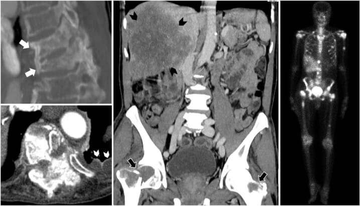
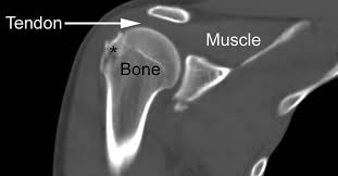
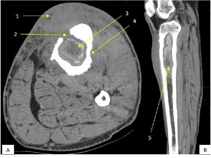
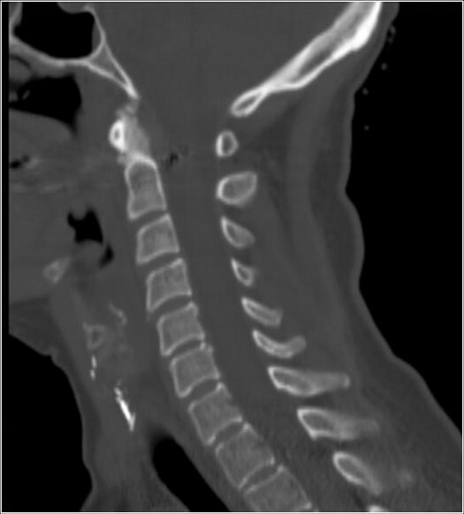
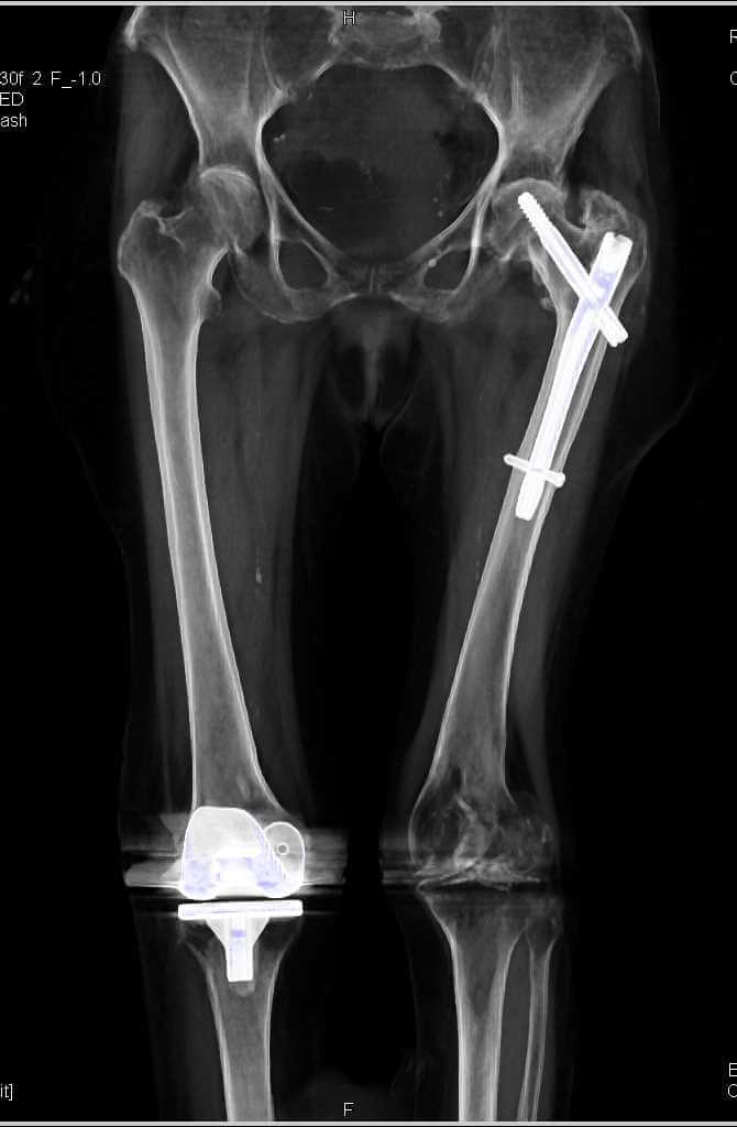
Sample bone CT scan images
Final word on bone CT scan and turn of bone CT scan
Bone CT scan is one of the best diagnostic imaging methods in the field of evaluation of damages to the skeleton, bones and masses of cancer. This scan is a procedure with and without injections. Keep in mind that if you want to go to the CT scan center to do a bone CT scan, be sure to have one with you. Try to get a bone CT scan from the nearest
CT scan
center.
To do this, you can easily book a CT scan from the nearest CT scan center. In a medical scan, you can find out exactly how to perform a bone CT scan, take turns CT scans of bones and see the answer to your bone CT scan.