
Interpretation of Pet Scan's answer
انتشار: 1401/05/20
Pet scan is one of the most powerful tools doctors have for diagnosing and monitoring the disease. Pet scans are often used along with CT or MRI, helping radiologists differentiate between healthy tissue and patient tissue so that cancer is accurately diagnosed or proper timing and treatment is performed. But for many patients and their loved ones, the complexity of pet scans can be a challenging experience. To better understand how Pet Scan works and its benefits, we're going to let you know the basic concepts of Pet Scan and to some extent teach you how to interpret the pet scan answer. So follow this article with us.
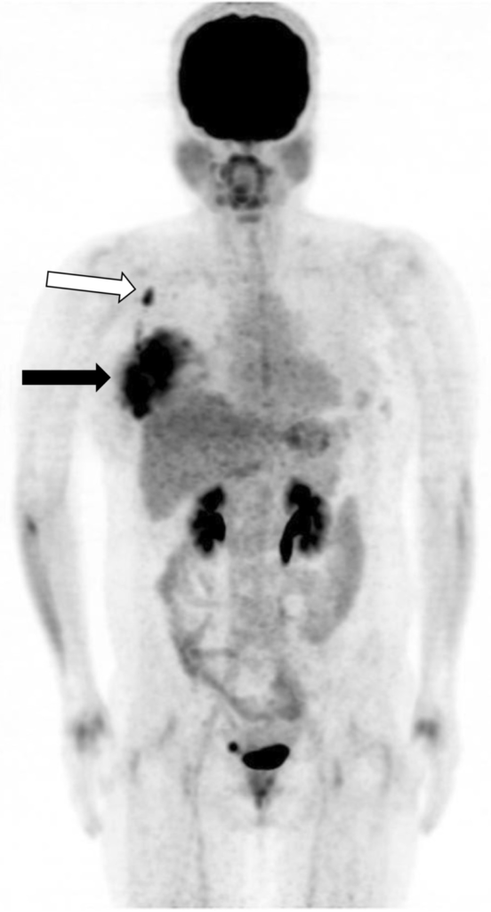
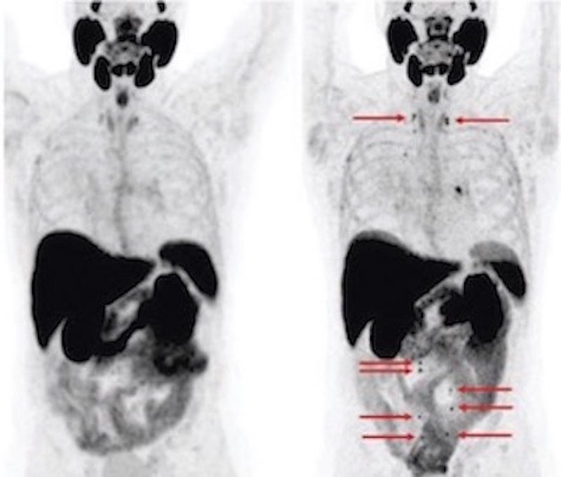 For many patients who receive pet scans, their number one question is about understanding the meaning of FDG absorption. Whether written in their pet scan response report is "non-absorption of FDG," "abnormal absorption of FDG," "low-grade FDG absorption" or any other changes - usually no explanation is mentioned in the report to help patients understand what this means.
FDG absorption refers to the amount of radiodharmuge absorption in body tissues. There is a perception among patients that any absorption of radiopharmaceevus is abnormal. However, this is not always true and can cause unnecessary alarm and concern.
So while a patient can take their report to their cancer specialist to explain their pet scan response, it is also curious to find out the results of their pet scan beforehand.
The Pet Scan report will also show another parameter called SUV (standard absorption value). The SUV is a benchmark used for regulatory purposes to see how things change over time. People don't necessarily have to worry about it, even if the numbers change dramatically. The SUV changes are supposed to be interpreted as part of the overall picture of what's going on.
In a CT scan, suppose you have a 1cm tumor and it doubles in size - now it all depends on the type of cancer and the type of treatment, but for the most part, people say yes - it shows me that the disease progressed.
Likewise, in a scenario where the intensity of a pet-scan SUV doubles over time, it is very common to assume that the disease has progressed. But actually, it's not necessarily always that way. The six-month changes should be a general assessment. Not just whether the numbers are increasing or decreasing. The amount of SUV is one of several ways to follow the scan, but this parameter is not an absolute way to check the meaning of the results.
For many patients who receive pet scans, their number one question is about understanding the meaning of FDG absorption. Whether written in their pet scan response report is "non-absorption of FDG," "abnormal absorption of FDG," "low-grade FDG absorption" or any other changes - usually no explanation is mentioned in the report to help patients understand what this means.
FDG absorption refers to the amount of radiodharmuge absorption in body tissues. There is a perception among patients that any absorption of radiopharmaceevus is abnormal. However, this is not always true and can cause unnecessary alarm and concern.
So while a patient can take their report to their cancer specialist to explain their pet scan response, it is also curious to find out the results of their pet scan beforehand.
The Pet Scan report will also show another parameter called SUV (standard absorption value). The SUV is a benchmark used for regulatory purposes to see how things change over time. People don't necessarily have to worry about it, even if the numbers change dramatically. The SUV changes are supposed to be interpreted as part of the overall picture of what's going on.
In a CT scan, suppose you have a 1cm tumor and it doubles in size - now it all depends on the type of cancer and the type of treatment, but for the most part, people say yes - it shows me that the disease progressed.
Likewise, in a scenario where the intensity of a pet-scan SUV doubles over time, it is very common to assume that the disease has progressed. But actually, it's not necessarily always that way. The six-month changes should be a general assessment. Not just whether the numbers are increasing or decreasing. The amount of SUV is one of several ways to follow the scan, but this parameter is not an absolute way to check the meaning of the results.
 The role of the referring physician is also discussed here. They must provide any laboratory work or other scans performed, as well as general clinical information about how the patient feels, to specialist radiologists.
Pet scans have great potential to guide patients and their doctors in the diagnostic and management stages of cancer. But the exact interpretation of PET scans is complicated - even more so than other types of imaging. In pet scans everything is not as clear and straightforward as CT or MRI.
Our advice is to make sure all your medical imaging is interpreted by the relevant specialist. There are many radiologists who start reading pet scans after taking a short training course. Although it may be enough for some cases, their knowledge base is not comparable to that of a nuclear medicine specialist.
To ensure accurate diagnosis and get the most accurate guidance on treatment response and cancer management – it's important to make sure you have a radiology specialist who interprets your pet scan.
Understanding the basics: How a pet scan works
The role of the referring physician is also discussed here. They must provide any laboratory work or other scans performed, as well as general clinical information about how the patient feels, to specialist radiologists.
Pet scans have great potential to guide patients and their doctors in the diagnostic and management stages of cancer. But the exact interpretation of PET scans is complicated - even more so than other types of imaging. In pet scans everything is not as clear and straightforward as CT or MRI.
Our advice is to make sure all your medical imaging is interpreted by the relevant specialist. There are many radiologists who start reading pet scans after taking a short training course. Although it may be enough for some cases, their knowledge base is not comparable to that of a nuclear medicine specialist.
To ensure accurate diagnosis and get the most accurate guidance on treatment response and cancer management – it's important to make sure you have a radiology specialist who interprets your pet scan.
Understanding the basics: How a pet scan works
 When a radiologist studies CT scans, radiology or MRI for diagnosis and staging of the disease, what the radiologist is looking for is something that looks different in shape, size, etc. While this assessment allows the doctor to diagnose abnormalities or physical changes, these abnormalities do not necessarily tell the doctor how they behave, while this is a very important part of assessing the disease. This is where the nuclear scan foot, especially the pet scan, opens up to the story.
Nuclear medicine imaging uses small amounts of radioactive material (called trackers) to examine the physiology (how) cells, molecules, chemical interactions, etc. are present in the body. For cancer and disease diagnosis, the most common nuclear scan is pet scan.
Most pet scan devices come with a CT scanner. This combination allows anatomical images of CT scans to be taken simultaneously along with pet images.
When a radiologist studies CT scans, radiology or MRI for diagnosis and staging of the disease, what the radiologist is looking for is something that looks different in shape, size, etc. While this assessment allows the doctor to diagnose abnormalities or physical changes, these abnormalities do not necessarily tell the doctor how they behave, while this is a very important part of assessing the disease. This is where the nuclear scan foot, especially the pet scan, opens up to the story.
Nuclear medicine imaging uses small amounts of radioactive material (called trackers) to examine the physiology (how) cells, molecules, chemical interactions, etc. are present in the body. For cancer and disease diagnosis, the most common nuclear scan is pet scan.
Most pet scan devices come with a CT scanner. This combination allows anatomical images of CT scans to be taken simultaneously along with pet images.
What does your pet scan answer say? (FDG Uptake, SUV)
For many patients who receive pet scans, their number one question is about understanding the meaning of FDG absorption. Whether written in their pet scan response report is "non-absorption of FDG," "abnormal absorption of FDG," "low-grade FDG absorption" or any other changes - usually no explanation is mentioned in the report to help patients understand what this means.
FDG absorption refers to the amount of radiodharmuge absorption in body tissues. There is a perception among patients that any absorption of radiopharmaceevus is abnormal. However, this is not always true and can cause unnecessary alarm and concern.
So while a patient can take their report to their cancer specialist to explain their pet scan response, it is also curious to find out the results of their pet scan beforehand.
The Pet Scan report will also show another parameter called SUV (standard absorption value). The SUV is a benchmark used for regulatory purposes to see how things change over time. People don't necessarily have to worry about it, even if the numbers change dramatically. The SUV changes are supposed to be interpreted as part of the overall picture of what's going on.
In a CT scan, suppose you have a 1cm tumor and it doubles in size - now it all depends on the type of cancer and the type of treatment, but for the most part, people say yes - it shows me that the disease progressed.
Likewise, in a scenario where the intensity of a pet-scan SUV doubles over time, it is very common to assume that the disease has progressed. But actually, it's not necessarily always that way. The six-month changes should be a general assessment. Not just whether the numbers are increasing or decreasing. The amount of SUV is one of several ways to follow the scan, but this parameter is not an absolute way to check the meaning of the results.
The role of the referring physician is also discussed here. They must provide any laboratory work or other scans performed, as well as general clinical information about how the patient feels, to specialist radiologists.
Pet scans have great potential to guide patients and their doctors in the diagnostic and management stages of cancer. But the exact interpretation of PET scans is complicated - even more so than other types of imaging. In pet scans everything is not as clear and straightforward as CT or MRI.
Our advice is to make sure all your medical imaging is interpreted by the relevant specialist. There are many radiologists who start reading pet scans after taking a short training course. Although it may be enough for some cases, their knowledge base is not comparable to that of a nuclear medicine specialist.
To ensure accurate diagnosis and get the most accurate guidance on treatment response and cancer management – it's important to make sure you have a radiology specialist who interprets your pet scan.
Understanding the basics: How a pet scan works
When a radiologist studies CT scans, radiology or MRI for diagnosis and staging of the disease, what the radiologist is looking for is something that looks different in shape, size, etc. While this assessment allows the doctor to diagnose abnormalities or physical changes, these abnormalities do not necessarily tell the doctor how they behave, while this is a very important part of assessing the disease. This is where the nuclear scan foot, especially the pet scan, opens up to the story.
Nuclear medicine imaging uses small amounts of radioactive material (called trackers) to examine the physiology (how) cells, molecules, chemical interactions, etc. are present in the body. For cancer and disease diagnosis, the most common nuclear scan is pet scan.
Most pet scan devices come with a CT scanner. This combination allows anatomical images of CT scans to be taken simultaneously along with pet images.