Difference between lung CT scan and lung radiology
Imaging plays an essential role in evaluating the lungs anatomically and lung function. Chest imaging, although it pays attention to determining and diagnosing and monitoring the severity of the disease, follows many goals. If you are suffering from any type of chest disease, your pulmonologist will most likely recommend that you take a chest X-ray or chest CT scan. Both of these methods are useful for helping your doctor determine the source of your symptoms as well as determining the best treatment plan for your specific needs. In this paper, we discuss the difference between lung CT scan and lung radiology, common imaging methods used to evaluate lungs, early technical aspects of imaging, and the appearance of common pathologies.

Lung
The lungs are cone and placenta-like organs inside the breast cavity that conduct gas exchange. They are covered by visceral and parietal layers of breast sides separated by pleural space, which typically contains about 10-20 milliliters of liquid. The lungs are centered by diaphragm in the lower, lateral and thoracic sides of the side with mediasten.
Placement of the components around the lung
The trachea splits to the right and left of the main nyj, which enters the lung from the umbilical region. The right lung is usually divided into the upper, middle and lower lobes by large fissure (diagonal) and minor (horizontal). The left is divided by a large gap into the upper and lower lobes. Each lobe is served by a second-order nyg that comes from the division of the main stem nyg. The lobes are divided into pulmonary bronchial sections based on third or segmental bronchials. Terminal bronchiols are eventually divided into respiratory bronchiols, where alveoli begin to appear.
Blood in the lungs
The lungs have double blood. Oxygenated blood from the bronchial circulation with 2 vessels to the left and one to the right and blood without oxygen through the circulation that is done through the lungs. Oxygenated blood is transferred to the right atrium through four pulmonary veins. The right bronchial vein is drained into the azigus vein and the left vein into the hemiazigote vein or the upper intercostal vein.
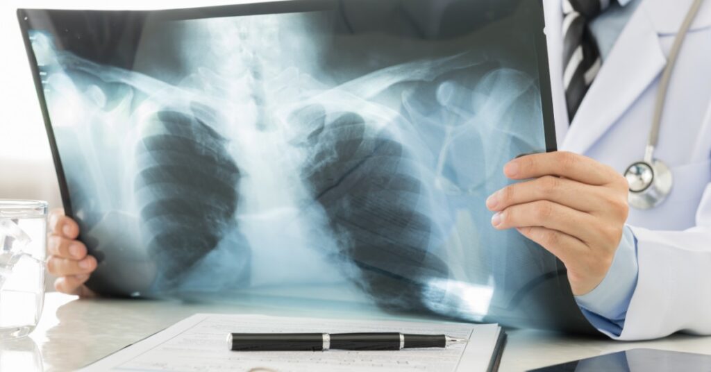
What is chest X-ray?
X-rays that use very small amounts of radiation in chest x-rays are one of the imaging methods that have a very good speed and high quality. This ray quickly passes through your body to get an inner image of your chest. Chest X-rays help show abnormal formations or a large variety of chest diseases such as pneumonia, cystic fibrosis, emphysema, cancer, etc. In addition, chest X-rays are often used to diagnose emergency diseases due to its fastness and quality.

What is a CT scan of the lung?
Chest CT scans are a more accurate type of chest X-ray that combines the power of X-rays and computers to provide a 3D view of the chest. A CT scan creates a number of precise incisions (images) that are then merged into one scheme. These images create a clear view that shows the position, shape and size of your breast organs.
Main differences in lung radiography and CT scan
1- Chest CT scan allows more details compared to chest X-rays. For example, chest X-rays may detect an abnormality, but chest CT scans should be able to show the exact location and examine the nature of the formation.
2- Chest X-rays provide a 2D image, while chest CT scans can create a 3D view of your organs.
3- X-rays are made to check for dense tissues, while better CT scans can record bones, soft tissues and blood vessels simultaneously.
4- X-ray equipment is much smaller and more complex than CT scan because the CT scan device should rotate around the scanned patient.
5- Chest X-ray is a low-cost examination with a good first appearance. To advance your diagnosis and treatment, you may have to do a chest CT scan to get a better picture.

Final Word
I hope we’ve been able to say some very useful things in this article so that now you can better understand the key differences between shooting with chest X-rays and chest CT scans. Don’t rush to decide which approach is most suitable for you. Remember, all cases are different and require an individual approach. Consult your doctor, he or she can answer any questions you have and prescribe the necessary tests for your specific conditions.


