All about radiology
All about radiology
Radiology or conventional radiography is a medical scan using X-rays. Conventional radiography (also called analog radiography, simple film or projection) is a basic diagnostic imaging tool in diagnosis and diagnosis of diseases. X-rays using the weakening or absorption of X-ray photons by high-density materials (such as calcium-rich bones) show differences in tissue structure. In this article, we are going to tell you all about radiology.

In conventional radiography, an X-ray beam is produced and transmitted to a footage or a radiation detector by passing through the patient’s body, producing an image. Different soft tissues vary X-ray photons depending on the density of the tissue. The denser the texture, the whiter the image (the clearer the beam). The range of densities, from highest to low density, is represented by metal (white or transparent), bone cortex (less white), muscle and liquid (gray), fat (darker gray), and air or gas (black, or black). Radiolucent).
Essentially, a conventional projection or radiography shows differences between bones, air and sometimes fat, which makes it particularly useful for assessing bone conditions and chest pathology. Low natural contrast between adjacent structures with similar radiographic density requires the use of contrast agents to increase contrast.
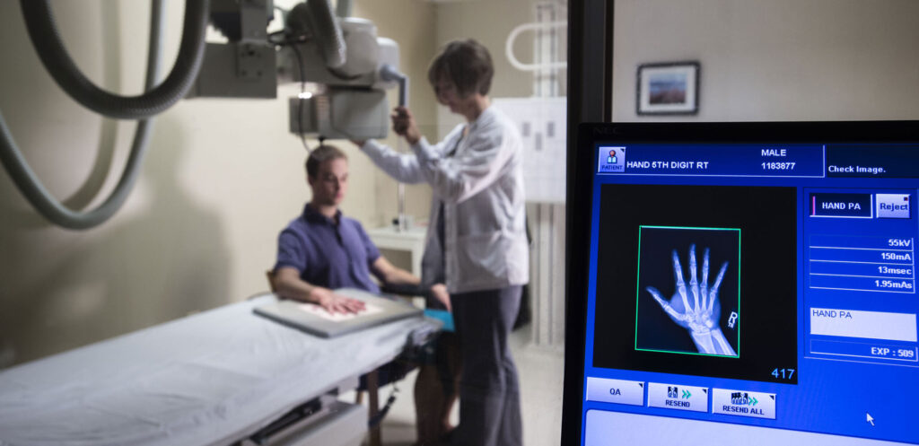
In conventional radiography, the patient is placed between an X-ray tube and a film or detector, sensitive to X-rays. The choice of the film and the resonant screen (which indirectly exposes the film) affect the resolution of the contrast and spatial resolution. Chemicals are required for film processing and are often the source of errors and re-harvesting. The result is a radiology scan, a still image that can hardly be manipulated after exposure to radiation. These images may also be viewed on fluoroscopic screens, movies or computer monitors.

X-rays are removed from the focal point of the X-ray tube as a divergent cone beam. For this reason, the radiographic design creates a variable degree of distortion. This effect decreases by increasing the source distance to the object relative to the distance from the object to the film, and using a collimator that passes only parallel X-rays.
The disadvantages of conventional radiography are lower contrast resolutions. Compared to computerized tomography (CT) and magnetic resonance imaging (MRI), it has the advantage of higher spatial resolution, inexpensive, easy to use and widely available. If the selected method is appropriate and sufficient, conventional radiography can yield high-quality results. X-ray systems and radioactive isotopes such as Iridium-192 and Cobalt-60 are also used to produce penetrating radiation in the body in non-destructive experiments.
Radiography is the most accessible imaging method. Typically, this radiology is the first imaging technique used to evaluate organs, chest and sometimes the spine and abdomen. These areas have important structures with different densities with adjacent textures. For example, radiography is the first scan to detect:
Fracture: In the image, bone radiology is seen as white because it is in the vicinity of soft gray tissues.
Pneumonia: In the radiology image, the inflammatory exudate, or pneumonia that fills the lungs, is well seen because it contrasts with adjacent airspace and a clearer beam.
Intestinal obstruction: Dilated, air-filled rings in the intestines are seen well among the surrounding soft tissue.
Radiology scan with contrast
When the density of adjacent tissues is similar, a contrast agent is often injected into a tissue or structure to differentiate it from its surroundings. Structures that usually require contrast agents include blood vessels (for angiography) and gastrointestinal lumina, biliary and urogenital tracts. Gas may be used to expand the lower part of the digestive system and make it visible.
Today, other imaging devices (such as CT, MRI) have largely replaced contrast radiology scans, as their 3D images provide a better anatomical image for an abnormality. Endoscopic procedures have largely replaced radiology scans with barium contrast in the esophagus, stomach and upper intestinal tract.
Fluoroscopy

Fluoroscopy is a subset of radiology devices, except that it shoots in real time and can come to the doctor’s aid during surgery. In fluoroscopy, a continuous X-ray beam is used to produce real-time images of moving limbs. Fluoroscopy is mostly used in the following cases:
- contrast agents (e.g., in swallowing or catheterization studies of coronary arteries)
- During medical measures to guide the placement of cardiac lead, catheter or needle (e.g. in electrophysiological tests or coronary interventions through the skin)
- Fluoroscopy can also be used in real time to detect diaphragm movement and bones and joints (e.g., to assess the stability of musculoskeletal injuries).
Disadvantages of conventional radiography
Diagnostic accuracy is limited in many situations. Other imaging tests may have advantages such as providing better or safer details or being faster.
Intestinal contrast agents such as barium and gastrographene (an iodine-based oral contrast agent) pose risks if used.
Fluoroscopy may include high doses of radiation.
Digital Radiography
Digital radiography, called DR, uses an X-ray-specific electronic detector that converts the radiographic image into a digital image to check on a computer monitor. The digital image is then stored and can be processed by changing the magnification, orientation, brightness and contrast. Digital radiography (also called direct radiography) is the progressive development of computerized radiography (CR).
The benefits of digital radiology can lead to lower radiation doses than analog or conventional radiography. DR and CR systems do not use any chemicals to process X-ray images, and hazardous materials and waste associated with film development are removed. Advantages of digital radiography compared to conventional radiography:
- Saves time and money due to more effective imaging process and workflow.
- Produce more quality images despite lower radiant doses.
- Lower repeat rates.
- Digital images can be stored on a disk or on a system as PACS images.
- Reduce dangerous chemical waste.
Final Word
Radiology is a very reich scan in medical imaging. To do a simple radiology, you need to spend a lot of time taking the turn of radiology. But medical scans provide a simple and free way to take the turn of radiology. You can also get many scans, such as ultrasound, CT scan, nuclear medicine, and MRI.


